Types of mammography and other breast imaging methods
August 17, 2024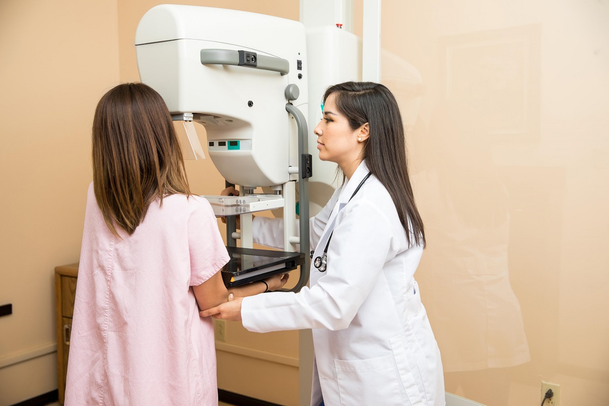
How is mammography performed?
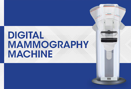
October 8, 2024Full field digital mammography Full-field digital mammography (FFDM), also known as “digital mammography” for simplicity, is a mammography system in which the film and cassette used in mammography with special plates called flat panels, similar to those in cameras, are replaced.
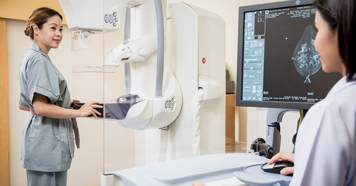
X-rays are converted into electrical signals, and then the electrical signals are used to produce images of the breast that can be viewed on a computer screen or printed on special films (similar to regular mammography films).
Types of digital mammography are:
direct radiography (DR or DDR), which is the most common type, and the image is formed directly on a flat panel.
Computed radiography (CR), which includes the use of a cassette containing an imaging plate
Digital breast tomosynthesis (DBT)
Mammography What is DBT?
Digital breast tomosynthesis is a relatively new technology. In DBT, the x-ray tube moves in an arc around the chest and takes several pictures from different angles. (such as computed tomography or CT scan), these images are then reconstructed into parallel “slices” of the breast. This allows interpreting physicians to see the overlapping layers of tissue separately.
Tomosynthesis (3D mammography): As mentioned in the previous paragraph, tomosynthesis, or “3D” mammography, is a new type of digital mammography with X-rays that creates two-dimensional and three-dimensional images of the breasts. This tool improves mammography’s ability to detect breast cancer early and reduces the number of women who are called back for additional tests (when their findings are actually non-cancerous).
“۳D” images reduce the overlap of breast tissue and allow the radiologist to better see your breast tissue on a mammogram.
Frequently asked questions about 3D digital mammography
Why is there a need for breast mammography by the tomosynthesis method? What are its benefits?
With a conventional digital mammogram, the radiologist gets a better view of the breast tissue, which is superimposed on the flat images. This tissue overlap sometimes makes it difficult to diagnose cancers. Also, the overlap can sometimes produce areas that look abnormal (false positives), so additional tests are needed to confirm that cancerous tissue is not present.
Tomosynthesis, or “3D” mammography, directly addresses the current limitations of standard 2D mammography. Several studies have shown that 3D mammography increases the detection of breast cancer by approximately 25% and reduces the number of false positive test results by approximately 15%.
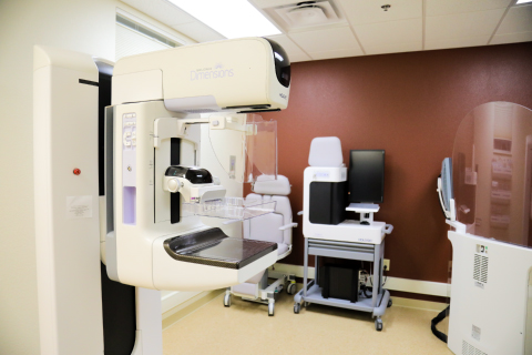
What should I expect during a 3D mammogram?
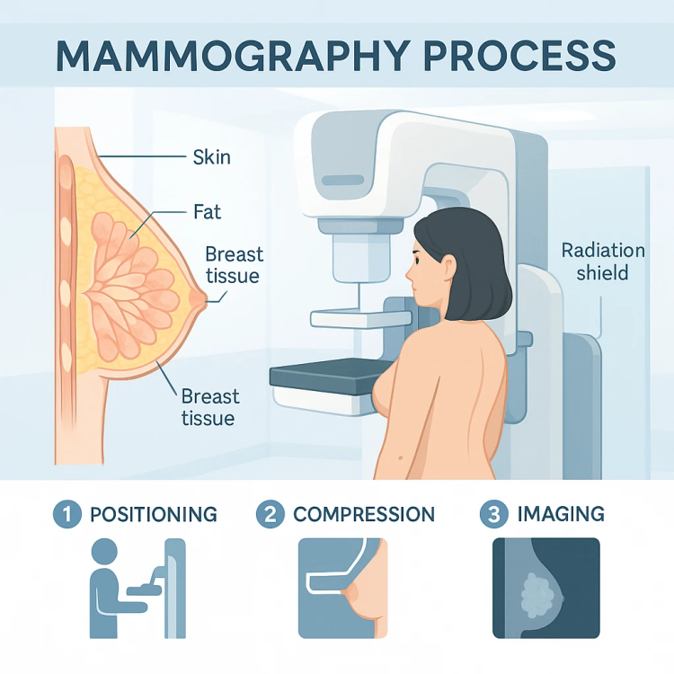
Performing a 3D mammogram is similar to a regular digital mammogram, including how much the breasts are compressed and how long it takes. The main difference is that the x-ray arm moves in a slight arc across your chest.
Computer-aided diagnosis
Computer-aided diagnosis systems (CAD) use a digital mammogram image that can be obtained from a conventional film mammogram or a digital mammogram. Computer software then looks for abnormal areas of density, mass, or calcification that may indicate cancer. The CAD system highlights these areas on the images and alerts the radiologist when further analysis is needed.
How can women find a digital mammography center?
Digital mammography has been used in the world since 2001 and can be performed in Iran as well. Some imaging centers in Iran still have analog mammography systems or first-generation digital mammography systems. CR) in which cassette is used. Golestan Radiology is proud to use advanced generations of digital mammography (DR), in which flat panels are used and images are interpreted by special mammography workstations.

