What is mammography?
August 17, 2024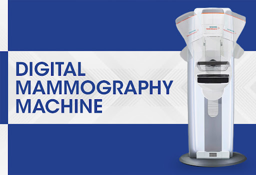
What is digital mammography?
October 8, 2024In this article, we are going to provide explanations about different types of mammography, including screening mammography, diagnostic mammography, two-dimensional mammography and three-dimensional mammography, as well as other methods of breast imaging. Mammography can detect breast cancer and track changes in your breasts over time, which helps doctors spot abnormalities and diagnose the disease in its early stages. It is very important to understand the different types of mammography, other types of available scans and the reasons for performing each one. In addition to screening and diagnostic mammography, there are other types of breast imaging, including MRI and ultrasound. Your doctor will decide which scan is right for you based on several factors.
How many types of mammography are there?
Annual screening mammography, which is used for early diagnosis in patients without clinical symptoms
Diagnostic mammography, which is used to detect suspicious cases in screening mammography or for patients with clinical symptoms.
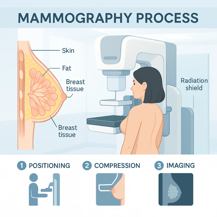
Screening mammography and diagnostic mammography can be two-dimensional or three-dimensional (also known as breast tomosynthesis).
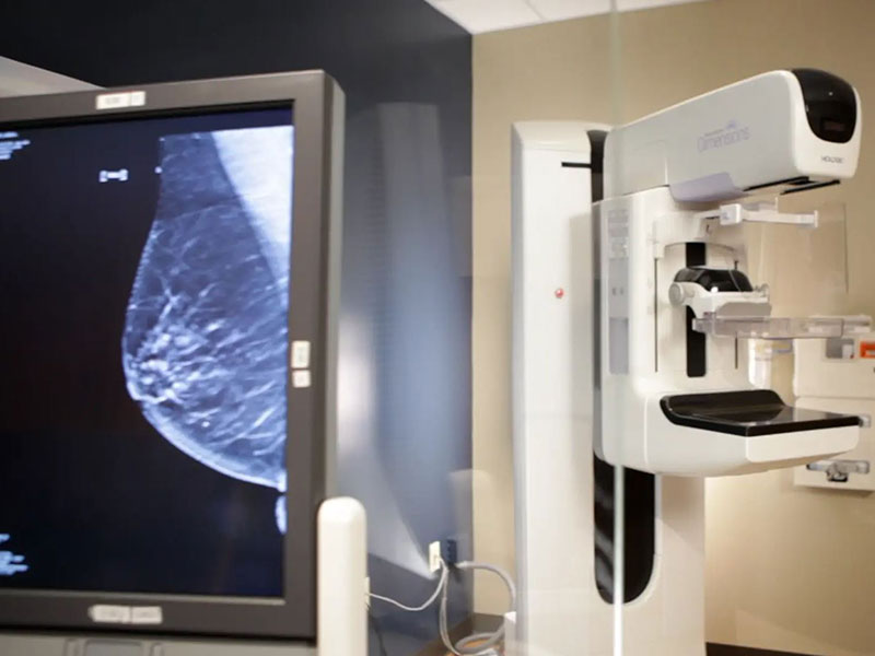
Screening mammography
It is used to detect breast cancer symptoms in women who have no symptoms.
X-ray images make it possible to detect tumors that cannot be felt.
It should be done annually at the age of 40 and older.
After the test, the radiologist interprets the images and finally informs the patients about the results.
The current test is compared to previous tests to find small anomalies.
Most of the time, the results are normal.
Diagnostic mammography
It is used to detect suspicious cases in screening mammography or when clinical symptoms such as lump, nipple pain, change in breast shape, nipple secretions, or change in color and orange skin of the breast skin occur.
Additional imaging such as ultrasound, MRI or biopsy may be required.
Two-dimensional mammography
Including digital mammography with flat and two-dimensional images
It can be limited due to overlapping layers of tissue that can sometimes produce inconclusive results, false alarms, or missing signs of cancer.
There is no need for additional compression (less pain), but compared to two-dimensional mammography, it takes a few seconds more for each display. The radiation dose received by the patient is slightly higher and it is more expensive than two-dimensional mammography.
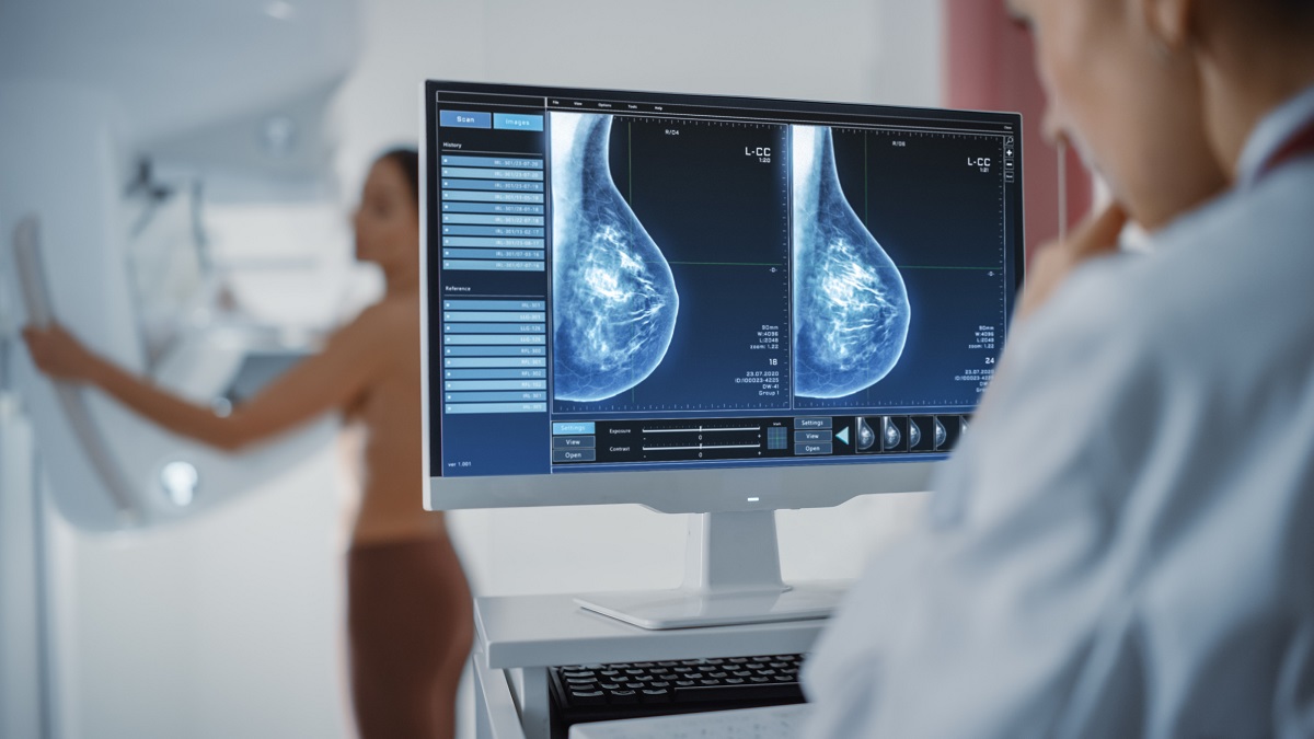
۳D mammography (tomosynthesis)
It is converted into a three-dimensional image using computer software. It allows the radiologist to examine the breast tissue layer by layer, and as a result, it has higher accuracy in breast cancer diagnosis.
This type is best for women with dense breast tissue, as both the dense breast tissue and the cancer appear white on a mammogram. By viewing the breast in layers, the radiologist can better differentiate the two.
No additional compression is needed (less discomfort), but it takes a few seconds longer per view compared to 2D mammography.
Can be more expensive than 2D mammography.
Breast MRI
For women who are at high risk of breast cancer or have dense breast tissue, often in addition to annual mammograms, a chest MRI is also recommended. MRI uses radio waves and a strong magnetic field to create detailed images of the inside of the breast. Mammography and breast MRI together can detect more cancers than using one method alone.
Differences between breast MRI and mammography:
But these two imaging methods have important differences. MRI alone is not recommended as a screening tool because this type of imaging can miss some cancers that mammography detects. MRI is also more likely to produce false positives. A false positive can lead to additional tests and biopsies that are not needed.
Doctors may advise some women under 40 to have an annual mammogram before starting. Do a chest MRI.
Women in this group may include those with a strong family history of breast or ovarian cancer or certain genetic mutations that increase the risk of breast cancer.
Faster results with rapid chest MRI
The last option for breast MRI is an innovative accelerated breast MRI. It is a highly sensitive imaging technique that shows more detail than digital mammography and takes less time than standard MRI, usually 12 to 18 minutes. This type of imaging is an accurate way to detect cancer in its early stages. In most cases, a rapid breast MRI requires the injection of contrast material to help radiologists see the details of certain structures and interpret the results more accurately. This scan can also be performed in an open MRI machine, providing more comfort for patients.
Breast ultrasound
Like breast MRI, breast ultrasound is not usually used alone as a screening method. But when a mammogram shows something unusual or a lump is felt in the breast, a breast ultrasound can give your doctor important information.
Often, doctors order a breast ultrasound to determine whether a lump is fluid-filled, which means it’s likely to be a benign cyst, or whether it’s solid. Lumps that are solid sometimes need a biopsy to determine if they are cancerous. The surgeon can also use ultrasound to help guide the biopsy needle to the correct area of the breast.
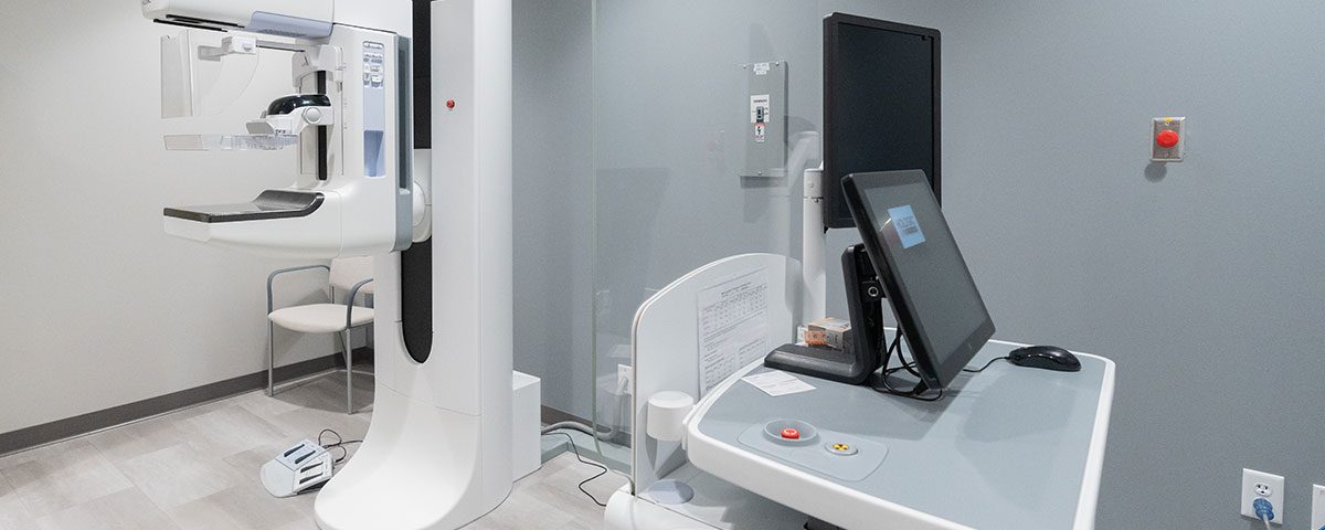
Some imaging centers in Iran still have analog mammography systems or first generation digital mammography systems, in which cassettes are used. Golestan Radiology is proud to use advanced generations of digital mammography (DR), in which flat panels are used and images are interpreted by special mammography workstations.
Also, the ultrasound machine of Golestan Center is equipped with an elastography system, which is one of the latest imaging technologies in determining the percentage of breast mass malignancy, and has been increasingly used by breast surgery specialists.
Automated breast ultrasound (ABUS) for additional tests
A technique called automatic breast ultrasound (ABUS) can be a good option for women with dense breast tissue. ABUS uses a much larger transducer to take hundreds of images that cover almost the entire breast. Doctors sometimes use ABUS for women who have abnormal mammograms or unusual symptoms.

