Types of mammography and other breast imaging methods
August 17, 2024Mammography is a specialized medical imaging technique that uses a low-dose X-ray system to see inside the breasts. Mammography helps early detection of breast diseases in women before they experience symptoms, when they are treatable.
X-rays help doctors diagnose and treat medical conditions by exposing you to a small dose of ionizing radiation to produce images of the inside of the body. X-rays are the oldest and most common form of medical imaging.
Three recent advances in mammography include digital mammography, computer-aided diagnosis, and breast tomosynthesis.
There are two types of mammography: screening mammography and diagnostic mammography.
What is a screening mammography?
A screening mammogram is a type of mammogram that is usually performed on women who have no signs or symptoms of breast cancer. Regular screening mammography can help reduce the number of deaths from breast cancer among women aged 40 to 74. It is important because it can detect breast cancer earlier and treat it earlier, before Its expansion begin.
But screening mammography can also have risks. Sometimes they can find something that looks abnormal but isn’t cancer. This leads to more tests and can cause you anxiety. Sometimes mammograms can miss cancer if it is exist. It also exposes you to radiation. You should discuss the pros and cons of mammography with your doctor. you can decide when and how often to have a mammogram.
What is diagnostic mammography?
A diagnostic mammogram is for people who have a lump or other signs and symptoms of breast cancer. These symptoms can include breast pain, thickening of the breast skin, discharge from the nipple, or a change in the size or shape of the breast. But these symptoms can also be caused by a benign breast disease (not cancer). Mammography with other tests, can help your doctor determine if you have breast cancer.

What is digital mammography?
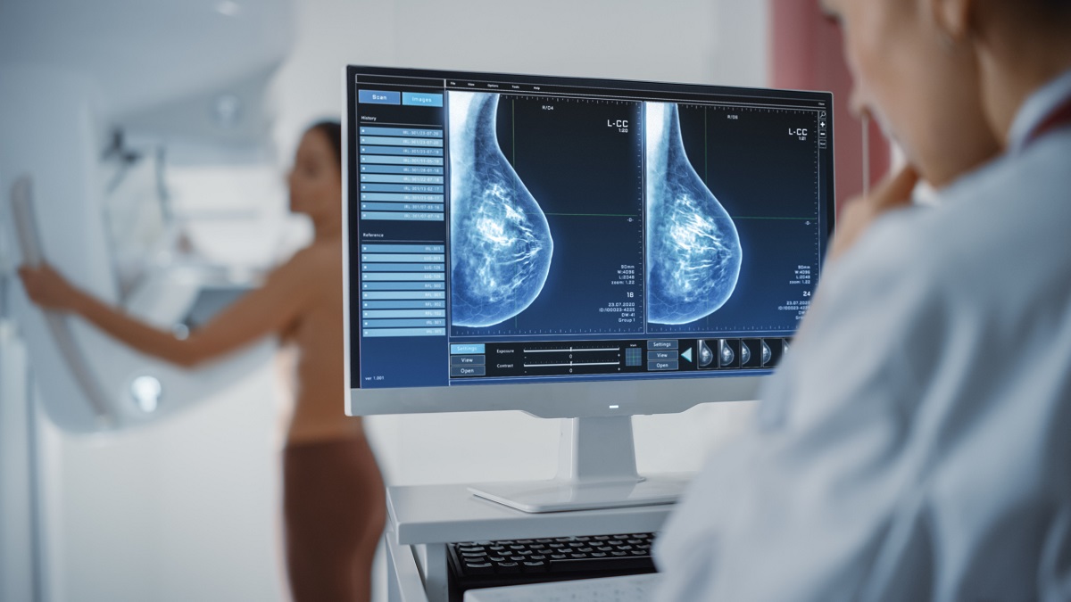
digital mammography which is called full field digital mammography(FFDM) is a mammography system which x-ray film is replaced by electronic devices that convert x-rays into mammographic images of the breast. These systems are similar to those found in digital cameras, and their efficiency results in better images with lower radiation dose. These breast images are transferred to a computer for review by a radiologist and for long-term storage. The patient experience during a digital mammogram is similar to conventional mammogram.
Some imaging centers in Iran still have analog mammography systems or first generation of digital mammography systems(CR) which cassette is used. Golestan Radiology Center is proud to use advanced generations of digital mammography(DR) in which flat panels are used and images are interpreted by special mammography workstations.
What is breast tomosynthesis mammography?
۳D mammography, also called digital breast tomosynthesis (DBT), is an advanced form of breast imaging which multiple images of the breast are taken from different angles and reconstructed (3D synthesis).
۳D breast imaging similar to computed tomography imaging (CT) which a series of “thin slices” are put together to create a three-dimensional reconstruction of the body.
Benefits of breast tomosynthesis:
Early detection of small breast cancers that may not be seen on regular mammography
Fewer unnecessary biopsies or additional tests
Possibility of diagnose multiple breast tumors
Clearer images of abnormalities in dense breast tissue
More accuracy in determining the size, shape and location of breast abnormalities
How is mammography performed?
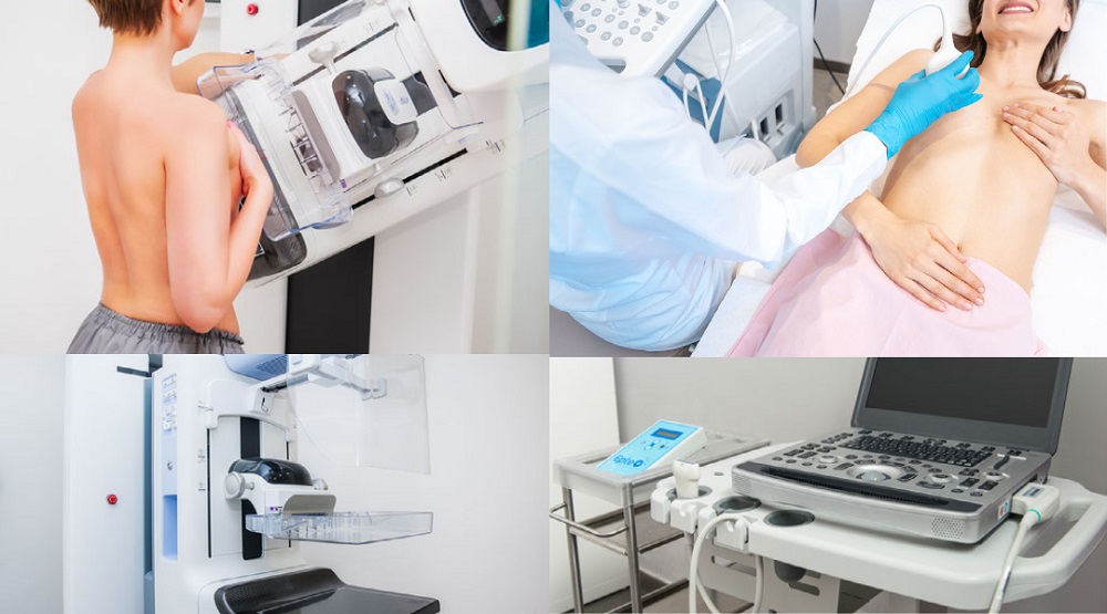
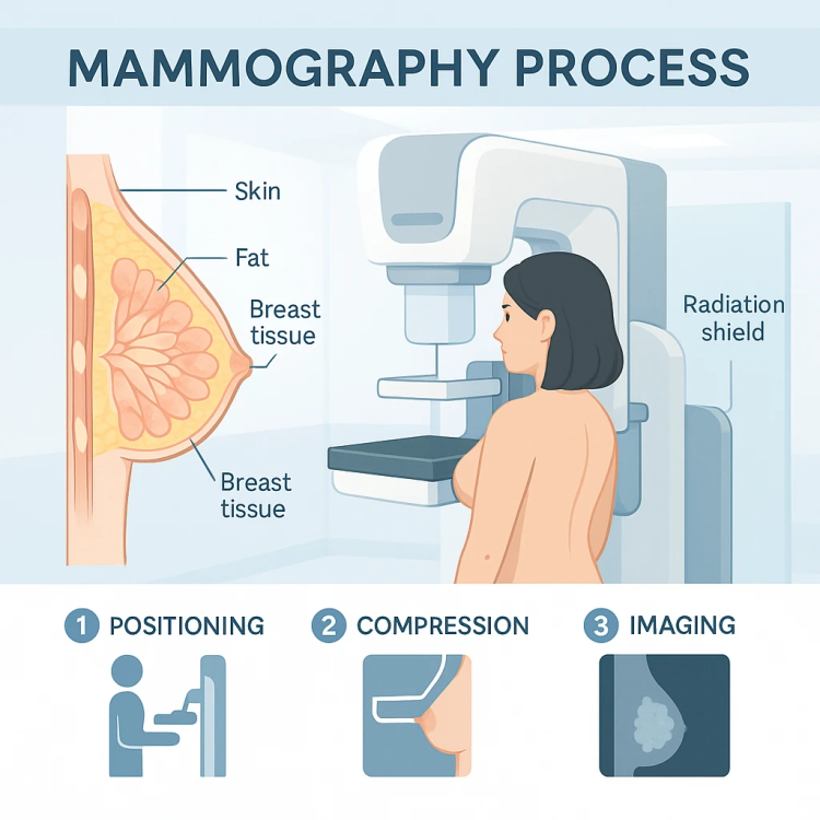
When you have a mammogram, you stand in front of an X-ray machine. The person taking the mammogram will place your breast between two plastic sheets. The plates compress and flatten your breast. This may be uncomfortable, but it helps to get a clear picture.
Both breasts are photographed from the front and from the side. Then the radiologist (doctor with special training) reads the mammogram. Your doctor will check the X-ray for early signs of breast cancer or other problems. You’ll usually get the results within a few weeks, although it depends on the clinic or doctor’s office you visited. If your results are not normal, you should start treatment sooner.

If my mammogram is abnormal, does that mean I have cancer?
An abnormal mammogram does not always mean cancer. You may need additional mammograms, tests, or examinations before your provider can make a diagnosis for sure. You may also be referred to a breast surgeon. But it doesn’t necessarily mean you have cancer or need surgery. You can go to one of these doctors because they are more specialized in diagnosing breast problems.
What are the benefits and risks of mammography?
Benefits of mammography
Screening mammography reduces the risk of death from breast cancer. To detect types of breast cancer, including invasive intraductal carcinoma (IDC) is useful.
Screening mammography improves the doctor’s ability to detect small tumors. When the cancers are small, the patient has more treatment options.
The use of screening mammography increases the detection of small abnormal tissue growth confined to the milk ducts in the breast, which is ductal carcinoma in situ (called DCIS).
After mammography (which is done with X-rays), no radiation remains in your body.
X-ray usually has no side effects in the normal diagnostic range in this method.
Risks of mammography
There is always a small chance of cancer from overexposure to radiation. However, due to the small amount of radiation used in medical imaging, the benefits of accurate diagnosis far outweigh the associated risks.
False positive mammography: 5 to 15% of screening mammograms require additional tests such as additional mammography or ultrasound. Most of these tests are normal. If there is an abnormal finding, biopsy may be needed. In false positive cases, biopsies confirm that cancer is not exist.
You should always tell your doctor and x-ray technician if you are pregnant.


