What is ultrasound
October 5, 2024
Types of pregnancy ultrasound
October 8, 2024Ultrasound is one of the most advanced methods in medical diagnosis, which is known as the safest imaging method. In this method, also known as ultrasound, ultrasonic waves are used, which cannot be heard by humans due to their high frequency.
The use of ultrasound led to a major breakthrough in medical science. Previously, imaging was mainly done with X-rays or doctors had to perform surgery to observe the inside of the patient’s body. Both methods had their own problems and risks.
Although X-ray imaging is still used in some special cases, due to its high sensitivity, it may damage the cells of the body if used incorrectly. Also, surgery without sufficient information about the internal condition of the body was considered a risky procedure. The discovery of ultrasound waves and the development of the ultrasound device solved these problems to a great extent. Ultrasound is used, which we will examine below.
What are the types of ultrasound?
Ultrasound is divided into different types according to the area being evaluated. In the rest of this article, we are going to explain each of these cases and make you more familiar with them.
External Ultrasound
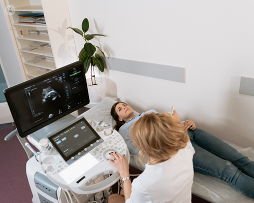
This type of ultrasound is performed on the body and is used for prenatal evaluations and examination of the fetal heart. The procedure is very simple: the doctor first covers the area with a special gel and then places the ultrasound probe on that area. By moving the probe on the surface of the body, the desired condition is checked.
Internal Ultrasound
This type of ultrasound is usually used to examine the prostate gland in men, as well as to evaluate the uterus and ovaries in women. To perform this ultrasound in men, the probe is inserted through the anus and in women through the vagina to provide the doctor with more detailed images.
Gastrointestinal and abdominal ultrasound
Abdominal ultrasound, a type of ultrasound used to evaluate the internal organs of the abdomen, such as the liver, spleen, gallbladder and pancreas. This method is not very effective for the detailed examination of the stomach and intestines, as empty spaces can interfere with the proper operation of ultrasound. However, in some cases it can be used to evaluate the vessels and walls of the stomach and intestines.
An ultrasound of the abdomen and pelvis can help diagnose:
- Lumps in the soft tissues or tears in the muscles of these areas
- Bleeding in the abdominal organs
- Infection and free fluid accumulation
- Identification of foreign body in the abdominal cavity
- Diagnosis of benign and malignant tumors in the abdominal and pelvic regions
- Evaluation of the kidneys and urinary tract
- Complete examination of the liver and ducts to diagnose problems such as liver cirrhosis, liver abscesses, and benign or malignant cysts
- Identifying inflammation associated with the gallbladder
- Detailed examination of the pancreas and diagnosis of pancreatic abscesses
Prenatal Ultrasounds
If a woman’s pregnancy progresses normally, her doctor will usually recommend four stages of ultrasound, which are planned as follows:
- Early pregnancy
- Week 12 of pregnancy
- Week 16 of pregnancy
- ۳۲ weeks of pregnancy

However, in some cases, for reasons such as the fetus being small or the mother suffering from diseases such as high blood pressure or diabetes, the doctor may recommend that the number of ultrasounds be increased or that some of them be repeated.
There are different types of prenatal ultrasounds that need to be performed using advanced devices. It is therefore advisable to visit imaging centers that have sourced their equipment from reputable sources.
- Transvaginal ultrasound
Vaginal ultrasound is one of the types of ultrasound that is performed during pregnancy on the recommendation of a doctor. This type of ultrasound differs from other methods, so that in this method, the probe of the ultrasound device is inserted into the vagina to examine the condition of the fetus, ovaries, uterus, and fallopian tubes. The vaginal allows for a more accurate assessment of the cervix.
- Ultrasound of the NT or back of the fetal neck
NT ultrasound is an imaging procedure performed between the 11th and 14th weeks of pregnancy. In this ultrasound, things like the fetus’s growth rate, heart rate, and brain development are examined. This type of ultrasound, which examines the back of the fetus’s neck, allows doctors to identify Down syndrome and other genetic defects. This ultrasound is performed in the abdomen and provides vital information about the health of the fetus.
- Fetal Health Ultrasound (Fetal Anomaly)
At 18 weeks of pregnancy, an anomaly ultrasound is performed to ensure the fetus’s complete health and growth in accordance with the gestational age. In this procedure, parameters such as the circumference of the head and the length of the longest bone in the fetus’s body are evaluated.
This type of ultrasound examines the fetus’s heart cavities, kidneys, bladder, brain, spinal cord, abdominal and sexual organs more closely. Also, it is necessary to evaluate the amount of amniotic fluid, check the fetal umbilical cord, placental position, and heart rate to confirm the complete health of the fetus.
- Four-dimensional ultrasound
A four-dimensional ultrasound provides the doctor with very precise details. If we want to explain the clarity of this ultrasound, we can say that even movements such as yawning of the fetus can be clearly seen in it. This ultrasound is usually requested by the doctor for further examination, and it is possible to perform it at any stage of pregnancy.
Endoscopic Ultrasound
Endoscopic ultrasound is an accurate method for examining some parts of the gastrointestinal tract that is performed in conjunction with endoscopic methods. This type of ultrasound can be done to examine the upper or lower parts of the gastrointestinal tract
In endoscopic ultrasonography of the upper parts of the gastrointestinal tract , the following are examined:
- Epithelial tissue of the wall of the esophagus, stomach and beginning of the small intestine
- Blood vessels and lymph nodes
- Other organs such as the liver, gallbladder and ducts
In endoscopic sonography of the lower parts of the gastrointestinal tract , the following are considered:
- Anal sphincter (anal valve)
- Blood vessels and lymph nodes, as well as cysts and tumors
- Epithelial tissue of the wall of the colon
Urinary tract ultrasound
This ultrasound is used to examine most components of the urinary tract, including the two kidneys, the ureters, the bladder, and the urethra. However, due to the small diameter and the urethra lying on top of each other, ureters cannot always be examined by ultrasound. Ultrasound of the kidneys and urinary tract can detect diseases such as enlargement of the kidneys, kidney stones, tumors, and other problems related to the urinary tract. It can be identified.
Cardiovascular Ultrasound
Cardiovascular ultrasound, also known as echocardiography, is used to diagnose abnormalities such as valve failure, improper blood pumping, and a general examination of the condition and function of the heart and its valves.
In this ultrasound, a two-dimensional incision of the heart is usually displayed, and if more advanced equipment is used, it is also possible to obtain a three-dimensional image of the heart.
Thyroid gland ultrasound
Thyroid ultrasound is one of the types of ultrasound that is performed to image the thyroid gland. This ultrasound may be requested for two purposes:
- Diagnosis of tumors and cysts: To identify and evaluate tumors and cysts present in the thyroid gland.2. Determine the position of the thyroid gland: To check the position of the thyroid gland, especially in cases where the gland may be stretched inward into the chest or behind the sternum.
Breast Ultrasound
Breast ultrasound is the first step in diagnosing breast cancer. This condition is very common among women, but men can also get it. If the treating physician suspects breast cancer, his first step is usually to refer the person to a breast ultrasound. This ultrasound is able to identify a variety of tumors and cysts in the breast tissue and provide important information for a more accurate diagnosis and treatment planning.

Eye Ultrasound
An ultrasound of the eye can provide useful information about the eyeball, the space behind it, the internal contents of the space behind the eye, and possible tumors in this area. This procedure helps the doctor to make a more accurate diagnosis of eye complications. People with eye problems, with the diagnosis of their treating doctor, should have an eye ultrasound to properly assess the health of their eyes.
Orthopedic Ultrasound
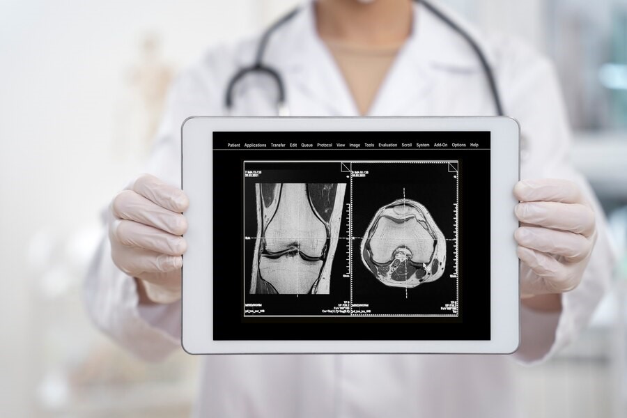
Orthopedic ultrasound is one of the types of ultrasound that examines the muscles, joints, tendons, and ligaments of the body. In this method, the patient is placed on a bed and the examined area is impregnated with gel so that the sonographer can perform the examination accurately.
Through orthopedic ultrasound, the following diseases can be diagnosed:
- Achilles tendon rupture
- Congenital hip dislocation
- Muscle tears in the shoulder area
- Knee meniscus injuries
- Internal muscle bleeding
- Tumors in the soft tissues of the body
Ultrasound in Gynecology
Due to the high importance of the female reproductive system, it is very important to examine it closely. With the advancement of ultrasound technology, it has become easier to diagnose various diseases in women, such as fibroids, uterine masses, ovarian cysts, and pelvic infections. Ultrasound allows doctors to identify many of these diseases at an early stage, which in turn can lead to more effective treatment and prevention. The progression of diseases.
Vascular Doppler Ultrasound
Doppler ultrasound is used to check blood flow in the veins. This type of ultrasound is able to identify various problems such as narrowing, blockage, and clogging of the arteries. Also, Doppler ultrasound helps to distinguish benign tumors from malignant ones and diagnose other vascular-related issues. This method provides doctors with accurate information about the speed and pattern of blood flow, which is very useful in evaluating and treating vascular diseases.
Women’s bladder ultrasound
Women may develop bladder diseases for a variety of reasons, such as childbirth and urinary tract infections. Bladder ultrasound is a useful tool for diagnosing these diseases. Ultrasound is used to assess the amount of urine remaining, the masses and stones in the bladder, and to measure its dimensions.
Also, when placing a catheter, an ultrasound of the bladder helps determine its exact position. For greater precision, a bladder ultrasound is usually performed first with a full bladder and then with an empty bladder.

Golestan Ultrasound proudly announces that it provides high-quality services to dear patients by using the most advanced equipment and the most up-to-date technologies, as well as relying on the knowledge and experience of a team of radiologists and skilled experts.
The center uses the latest ultrasound technologies, including liver elastography to diagnose diseases such as fatty liver, fibrosis, and liver cirrhosis. Also, breast and thyroid elastography is available, which is used to assess and determine the risk of malignancy in breast and thyroid masses.
In addition, Golestan Ultrasound Center strives to provide a safe and comfortable experience for its patients by providing a calm and professional environment. Our main focus is on providing quality services, high accuracy, and special attention to the needs and concerns of each patient. We are committed to making accurate diagnosis and appropriate treatment possible for our dear patients by using advanced technologies and up-to-date knowledge of the world.
Ultimately, our goal at Golestan Ultrasound is to ensure the health and satisfaction of patients. We proudly continue to provide specialized services in the field of advanced ultrasounds and are always striving to provide you with the best.


