Types of ultrasound
October 8, 2024Ultrasound is a diagnostic procedure that produces clear and accurate images of internal organs and tissues by sending high-frequency sound waves into the body. This method is especially important in the fields of obstetrics and gynecology.
The main purpose of ultrasound is to diagnose and image organs and body tissues in order to identify possible diseases and defects, such as problems in the uterus, ovaries, breast, liver, and other organs. In addition, ultrasound is also used to measure blood flow, identify inflammatory areas, and examine the fetus during pregnancy.
This method is frequently used because of its safety, especially for fetal evaluation in pregnant mothers. Ultrasound has a significant advantage over radiography and other methods that use X-rays because X-rays may be dangerous for the mother and the fetus.

Ultrasound can be performed in the following forms
۲D: It provides images of the inside of the body in a flat and two-dimensional way.
۳D: It adds an extra dimension to the images and provides 3D images of the internal structures of the body.
۴D: Combines 3D images with motion, and the result is displayed as a video or video.
How is ultrasound performed?
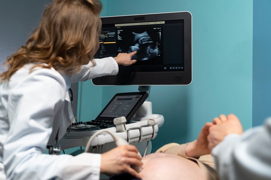
To perform an ultrasound, the person must lie on the bed. To improve the transmission of sound waves, the area is covered with a gel to prevent air from forming between the probe and the body. The doctor then moves the probe over the skin.
In this process, the doctor can view the backwaves through the ultrasound machine in the form of an image. The doctor’s skill in providing accurate and appropriate images and choosing the right angle for imaging is crucial.
The recorded images are immediately prepared and provided to the patient. The doctor also provides a report to the patient along with the images. Therefore, the doctor’s skill and accuracy of the ultrasound device are of great importance in providing a correct diagnosis. The ultrasound probe can be used both inside the body (such as vaginal ultrasound) and outside the body to visualize the internal organs.
What happened during the ultrasound?
Ultrasound is a medical imaging procedure that uses high-frequency ultrasound waves to produce images of the body’s interior. During this process, your doctor will tell you how to lie down or sit and explain other necessary tips.
You lie on the ultrasound bed and the area of the body to be examined is covered with ultrasound gel so that the ultrasound waves penetrate more effectively into the body and create higher quality images.
Then, using an ultrasound device, the doctor sends ultrasound waves to the area of interest. These waves penetrate deep into the body and image details of internal tissues and structures.
During this process, the doctor carefully examines and analyzes the resulting images. After the ultrasound is completed, the images are provided to the doctor so that he or she can make the necessary diagnoses about your health condition.
Benefits of Ultrasound
Ultrasound is not dangerous due to the absence of X-rays, unlike radiography. This method provides clear images of the internal organs of the body. It is also safe for pregnant mothers and fetuses and has many applications. In this technique, injections or invasive procedures are not used and the cost is relatively affordable. Also, ultrasound is done painlessly.
What is ultrasound used for?
Ultrasound is a medical imaging procedure that is used to diagnose and investigate various problems in the internal organs and tissues of the body. This method is usually used for:
- Cardiac ultrasound: to evaluate the structure, movement and function of the heart , vascular
- Abdominal ultrasound: to diagnose problems related to the liver, bile, kidney, pancreas and other abdominal organs
- Breast ultrasound: to identify and follow up breast tumors and check their location
- Ovarian ultrasound: to diagnose and follow up on problems such as cysts and ovarian infections
- Skin ultrasound: to check for skin problems such as infections
- Brain ultrasound: to diagnose brain problems such as bleeding or tumors
- Joint ultrasound: to identify joint problems such as inflammation and injuries
- Obstetric ultrasound: to monitor fetal development and uterine condition during pregnancy
- Orthopedic ultrasound: to diagnose problems such as joint injuries and soft tissue disorders.
How does ultrasonography or ultrasonication work?
Ultrasound uses high-frequency sound waves that are inaudible to the human ear. When these waves hit the internal organs of the body, they are reflected and interpreted by the ultrasound machine as an image.
The procedure is capable of imaging structures such as ligaments, tendons, arteries, and other tissues. Even when the body’s internal organs are moving, such as cardiovascular, ultrasound has the ability to provide clear images.
During pregnancy, this method is widely used to monitor fetal growth, identify possible fetal defects and problems. Also, ultrasound is very useful in detecting cancerous masses in the breast, thyroid gland, and other areas of the body.
Preparations for Ultrasound
In general, ultrasound does not require any special preparation, except in some special cases. For example, in a gallbladder ultrasound, it is necessary for a person to drink nothing but water for about 6 hours to get clear images of the gallbladder.
For ultrasound of the pelvis and its internal organs, such as the uterus and ovaries, the person needs to drink plenty of water to fill their bladder, which helps to get better images of the inner parts of the pelvis.

What are the things to do before having an ultrasound?
Before performing an ultrasound, paying attention to the following can help improve the quality of the results:
- Doctor’s guidance: Before performing an ultrasound, consult your doctor about any medications or medical conditions you have and follow his instructions carefully.
- Prepare documents: If you have had an ultrasound before, bring documentation of the previous results so that the doctor can compare the new results with the previous results.
- Diet: Before the ultrasound, it is better to consume only liquids (except milk) for about 6 hours or have a very light diet such as a little bread and jam (especially in diabetic patients who may face hypoglycemia).
- Pay attention to previous diseases: If you have diseases such as diabetes, consult your doctor before an ultrasound.
- Wear appropriate clothing: For comfort in performing ultrasounds, wear comfortable and appropriate clothing to make it easier to access the required areas.
Who interprets the ultrasound results?
Your radiologist will send ultrasound images to your doctor in the form of a medical report. By visiting your doctor, who has prescribed you an ultrasound, you can find out about your physical condition based on the results of the ultrasound.
Does it hurt to have an ultrasound?
No, ultrasound is completely painless, as it moves over the skin and captures images using only the device. For this reason, this procedure is not only painless, but also does not cause any discomfort. Therefore, there is no reason to worry or fear in this regard.

Conclusion
Ultrasound is an imaging method using sound waves that is able to provide images of the internal organs of the body. Due to its safety, this method is widely used in obstetrics and gynecology issues, but it should not be used continuously and in unnecessary situations.
A person can perform an ultrasound without stress, pain, or discomfort. With the presence of a skilled doctor and the use of a high-precision and high-quality ultrasound device, the results can usually be received at the same time.
Golestan Sonography is proud to provide suitable conditions for providing sonography services to patients by using advanced and up-to-date devices and relying on the knowledge and experience of the team of radiologists and experts.
In this center, the latest technologies used in sonography such as liver elastography (for diagnosis of fatty liver, fibrosis and cirrhosis of the liver), as well as breast and thyroid elastography (for determining the risk of malignancy in breast and thyroid masses) are available.


