Types of ultrasound
October 8, 2024
Everything You Need to Know About Pregnancy Ultrasound
October 8, 2024Seeing the newborn for the first time through a screen can be an emotional and overwhelming experience for the mother, but ultrasound is vital for several other reasons. Ultrasound can assist in monitoring the growth of the fetus, screening for potential issues, and overseeing the health of you and your child. But what are the different types of pregnancy ultrasounds, and when do you need them? Depending on the stage of pregnancy and also for various reasons, such as calculating the likelihood of the baby having Down syndrome, the risk of premature birth, and many other factors, different types of pregnancy ultrasounds are performed.
Types of pregnancy ultrasound scans
• Initial pregnancy scan (determining gestational age) (weeks 8 to 11)
• Nuchal translucency scan (NT) (weeks 11 to 14)
• Fetal anomaly scan from week 18 to 23.
• Growth scan or fetal health scan (weeks 24 to 42)
Initial scan (determining gestational age) (weeks 8 to 11)
The first ultrasound you may experience will be between weeks 8 to 14 of pregnancy to estimate the due date of the baby. This scan is performed to confirm pregnancy and determine the estimated due date (EDD). The fetal heartbeat is detected in this scan. Most parents choose this scan for peace of mind. Knowing this date is also essential for calculating the likelihood of Down syndrome through a non-invasive prenatal test (NIPT) conducted before a blood test. This scan is performed on your abdomen. Sometimes, it may be necessary to have an internal (vaginal) scan to obtain more information and a clearer picture.
Nuchal translucency scan (weeks 11 to 14)
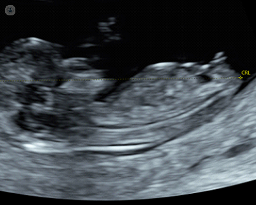
This scan is usually performed through the abdomen, and sometimes an additional internal scan may also be required. This scan is performed for the following cases:
• Confirmation that the child’s heart is beating.
• For a more accurate estimation of the duration of pregnancy.
• To measure the length of a newborn.
• To determine whether there is more than one baby (multiple births) and to assess whether there is a shared placenta, which requires close monitoring during pregnancy.
• To assess the structure of the fetus, including measuring the fluid at the back of the baby’s neck.
• Examining the likelihood of a newborn developing common chromosomal issues such as Down syndrome, Edwards syndrome, and Patau syndrome.
Fetal anomaly scan (weeks 18 to 23)
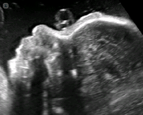
This scan is usually performed between weeks 18 and 23. This scan is a systematic examination of the newborn from head to toe so that the doctor can see if the structures appear normal as expected for this age. In this scan, most abnormalities are detected if present.
During this scan, the position of the placenta and blood flow through the placenta will also be assessed. In addition, the risk of preterm labor can be predicted by measuring the length of the cervix with an internal scan.
Growth scan from week 24 onwards
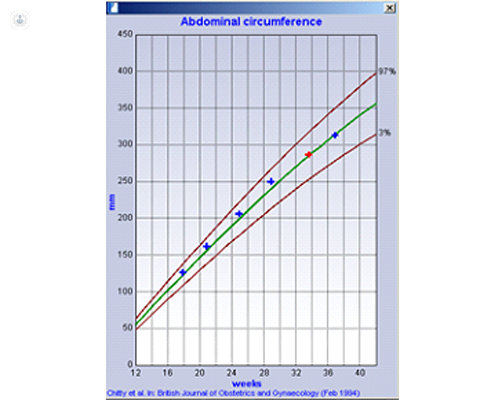
The growth and health of the newborn after 24 weeks are monitored by these scans.
Specific measurements of the head, abdomen, and thigh of the newborn are taken to estimate the weight of the fetus.
The volume of amniotic fluid surrounding the newborn is measured.
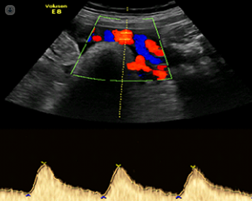
Blood flow is examined through the umbilical cord and the blood vessels of the newborn. (Doppler scan for newborns)
Types of technologies used in pregnancy ultrasounds
Standard two-dimensional scans
The standard ultrasound test performed by hospitals and clinics is two-dimensional. A standard two-dimensional ultrasound produces flat and clear images that can be used to view your child’s internal organs and assist in diagnosing any internal issues.These black-and-white images help the sonographer determine the pregnancy, growth, heartbeat, development, and size of the baby.
۳D scans
Some pregnant mothers choose 3D ultrasound scans because they provide a more realistic image of the baby. You can have a 3D scan between weeks 26 and 30 of your pregnancy; this scan allows your doctor to see a detailed 3D image of the fetus.
Four-dimensional scans
۴D ultrasound scans are similar to 3D scans, but instead of static images, they show a moving video. This work better showcases the shadows and highlights, thus creating a clearer image of the child’s face and movements.
Both 3D and 4D ultrasound scans can be performed from the 26th week of pregnancy.
Transvaginal ultrasound
Transvaginal ultrasound is used to examine internal structures. This test is performed with a small probe that is inserted into the vagina and is usually used in the early stages of pregnancy, when obtaining a clear image from the outside is difficult.
Points of all ultrasounds.
All types of ultrasound are safe and low-risk. It may be done for medical and non-medical reasons, but unnecessary exposure to ultrasound waves is not recommended according to medical guidelines. The average number of ultrasounds during each pregnancy varies and depends on the results of your tests and initial scans.

