Types of pregnancy ultrasound
October 8, 2024
The difference between mammography and ultrasound
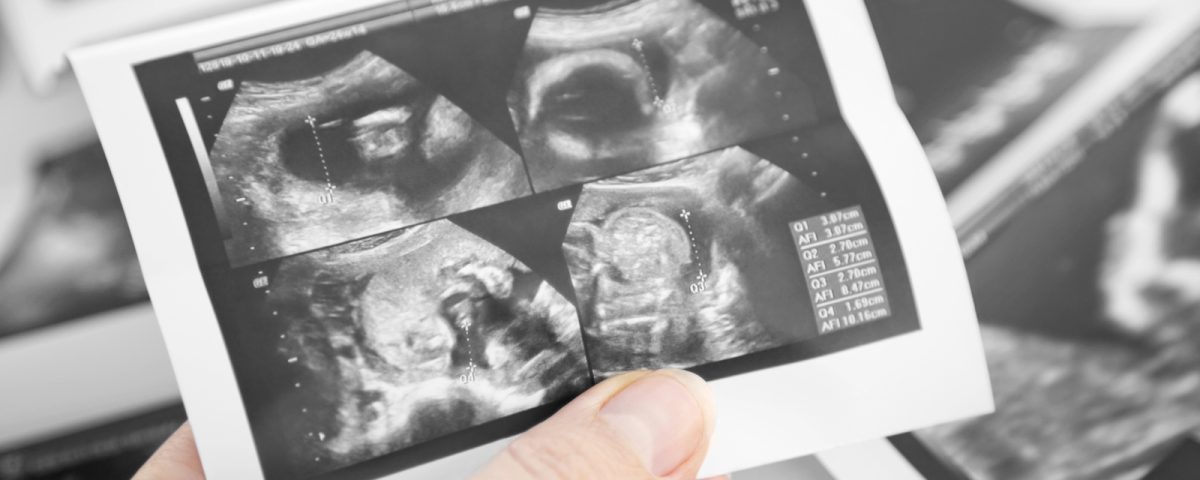
October 8, 2024Pregnancy ultrasound is an imaging test that uses sound waves to create an image of how the fetus is developing in the womb. This test is also used to examine the pelvic organs of women during pregnancy. As mentioned, ultrasound during pregnancy is used to assess the health of the fetus and to diagnose certain pregnancy complications.
The number of ultrasounds during pregnancy depends on the conditions of the mother and fetus, as well as the doctor’s diagnosis, and it varies slightly in different countries.
What is a pregnancy ultrasound?
It is a test during pregnancy that examines the health and growth of your child.
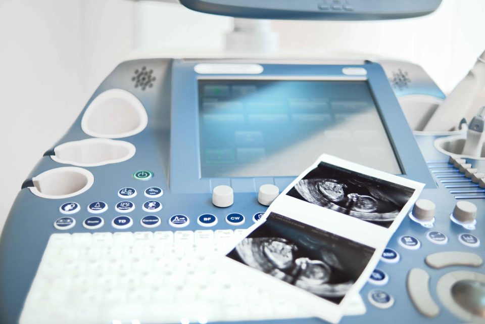
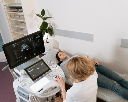
During the ultrasound, sound waves are transmitted by a device called a transducer (probe) through the abdomen or vagina. Sound waves are reflected from the structures inside your body, including the fetus. Then, sound waves are converted into images that a specialist can see on the screen. In this method, rays such as X-rays are not used.
Although pregnancy ultrasounds are safe, they should only be performed when necessary. If there is no reason for an ultrasound (for example, if you just want to see your baby), it is recommended not to have the ultrasound done.
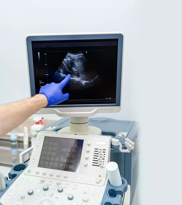
What can be diagnosed with a pregnancy ultrasound?
A pregnancy ultrasound serves two purposes:
It assesses the overall health, growth, and development of the fetus.
It diagnoses some diseases and medical issues related to pregnancy.
The reasons for performing a pregnancy ultrasound include:
• Pregnancy confirmation
Examination of ectopic pregnancy, molar pregnancy, miscarriage, or other early pregnancy risks.
• Determining the start time of pregnancy and establishing the due date.
• Examining the growth, movement, and heartbeat of the fetus.
Determining twins, triplets, or more!
• Examination of pelvic organs such as the uterus, ovaries, and cervix.
• Examination of the amount of amniotic fluid
• Examination of the mating site
• Examination of the fetus’s position in the womb.
• Diagnosis of problems related to fetal abnormalities.
When is the first ultrasound during pregnancy performed?
The timing of the first ultrasound varies depending on your and the doctor’s opinion. For example, some individuals undergo the initial ultrasound, which takes place in the early weeks of the seventh to eighth week of pregnancy.
Some initial ultrasound examinations are performed via the vagina (transvaginal ultrasound), which has greater accuracy for observing the gestational sac, the fetus, and the fetal heartbeat, especially if an ultrasound is needed between the fifth and seventh weeks (because the gestational sac and fetus are smaller, or the heartbeat may be questionable through abdominal ultrasound). This method has higher accuracy.
The initial ultrasound determines the following:
• Confirmation of pregnancy (with detection of heartbeat).
• Determining whether there are multiples or not.
• Measuring the size of the fetus
• Determining the time of pregnancy and the due date.

Ultrasound at the twentieth week (anatomy scan)
An anatomical ultrasound is performed around weeks 18 to 20 of pregnancy.
During this ultrasound, some anatomical abnormalities of the fetus, such as serious issues related to the brain, heart, bones, or kidneys of the child, are diagnosed. If your pregnancy is going well and without complications, the ultrasound at week 20 may be your last ultrasound during the pregnancy.
Additionally, in this ultrasound, you can find out the gender of your child (if the baby is in a suitable position to observe the genitalia).
Main types of pregnancy ultrasound
There are two main types of pregnancy ultrasound: transvaginal ultrasound and abdominal ultrasound. Both use the same technology to produce images. Transvaginal ultrasound is performed by inserting a special probe inside the vagina, while abdominal ultrasound is conducted by placing a device on the skin of the abdomen.
Standard ultrasounds are two-dimensional. Advanced technologies such as ultrasound image reconstruction can generate additional information by producing three-dimensional or four-dimensional images. This type of ultrasound is useful when the doctor needs to see the face or limbs of the fetus in greater detail. Not all clinics have 3D or 4D ultrasound equipment or trained and specialized personnel to perform this type of ultrasound.
Your provider may recommend other types of ultrasound. Examples of additional ultrasounds include:
The above sentence is incomplete and does not continue.
First-trimester screening ultrasound (NT) to determine the risk of chromosomal abnormalities.
Doppler ultrasound of the uterine vessels, umbilical cord, and fetus to assess blood supply to the fetus.
Biophysical ultrasound for respiratory movements, body movements, and tone or degree of fetal body contractions.

Getting ready for a pregnancy ultrasound
Some centers may recommend having a full bladder (this is more helpful during weeks five to seven).
You may be asked to drink 2 to 3 glasses of fluids an hour before the test. Also, you should not urinate before the test.
Pressure on the bladder may cause discomfort. The conductive gel may feel a bit cold and damp. You will not feel the ultrasound waves.
Pregnancy ultrasound results
Normal or regular results
The growth of the fetus, the placenta, the amniotic fluid, and the surrounding structures appears to be normal.
Note: Normal results may vary slightly. Talk to your doctor about the meaning of your specific test results.
Unusual results
Abnormal ultrasound results may be due to some of the following conditions:
• Congenital defects
• Ectopic pregnancy
• Weak fetal growth in the mother’s womb.
• Multiple pregnancies
• Miscarriage
• Problems with the child’s position in the womb.
• Pair problems
- Very low amniotic fluid
• Excess amniotic fluid (polyhydramnios)
• Pregnancy tumors, including trophoblastic disease.
• Other issues related to the ovaries, uterus, and pelvic structures.
The dangers of ultrasound
According to current research, current ultrasound techniques are safe. Ultrasound does not involve radiation.

