What is radiology
October 5, 2024
tests and examinations related to pregnancy
October 8, 2024Types of Medical Imaging and the Application of Each
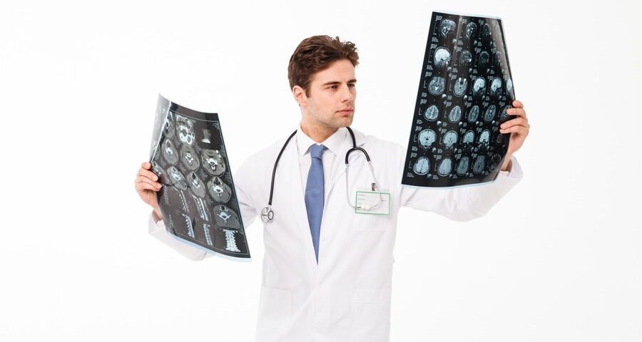
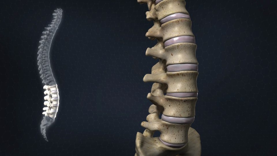
Radiology plays a key role in diagnosing and treating diseases by using various medical imaging techniques. This field includes a variety of imaging methods, each with its own features and benefits. In this article, we will explore the different types of radiology and the unique applications and benefits of each.
The importance of radiology
Radiology is a vital part of modern healthcare, allowing doctors to accurately observe the internal structures of the body. These techniques provide valuable diagnostic information that helps diagnose, evaluate, and track various medical conditions. The various radiology techniques are each designed for specific purposes, and as such, they have become valuable tools for medical professionals.
Types of Medical Imaging
-
X-ray radiology
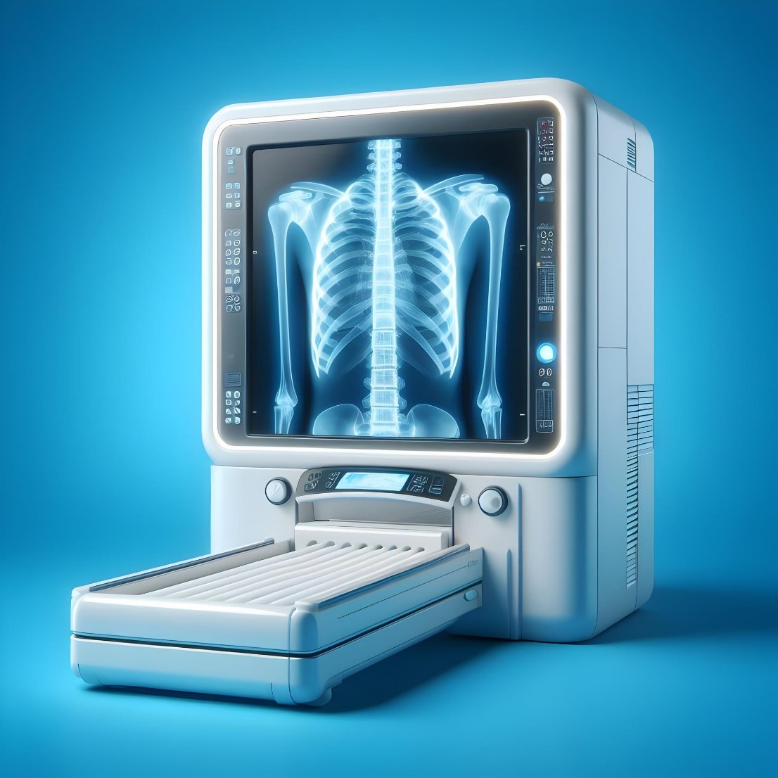
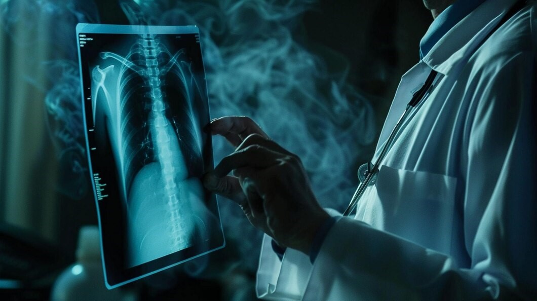
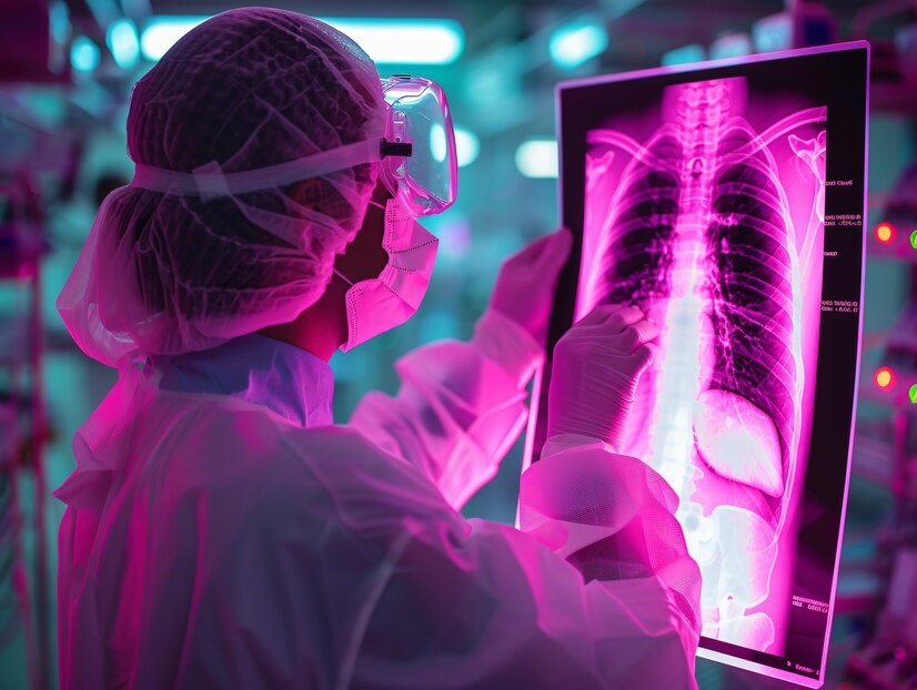
X-ray imaging is one of the oldest and most widely used methods of medical imaging. This technique involves the use of electromagnetic radiation that is used to create images of bones, tissues, and internal organs of the body. X-rays are particularly effective in detecting fractures, infections, tumors, and abnormalities of the skeletal system. In dental cases, a CBCT scan is used, which provides a three-dimensional image of the patient’s jaw and face.
-
Computed tomography scan (CT scan)
Computed tomography (CT) is an advanced imaging procedure that combines X-rays and computer technology to provide accurate cross-sectional images of the body. This technique helps medical professionals obtain a more comprehensive view of the body’s internal structures and accurately diagnose and describe various conditions.
In the CT scan process, the patient lies down on a table that passes through a circular ring in the CT machine. The machine shines X-ray beams around the body from different angles. The detectors measure the amount of X-rays passing through the body and convert it into digital signals. These signals are then processed by a computer and cross-sectional images or slices It is produced precisely from the body.
Because of its ability to visualize the exact details of bone structures and soft tissues, CT scans are a very effective tool in diagnosing a wide range of medical conditions. The resulting images can show abnormalities such as tumors, infections, blood clots, or internal damage.
The high accuracy of CT scans in providing cross-sectional images allows doctors to examine structures layer by layer, providing a more comprehensive picture of internal anatomy. This feature is especially useful in evaluating complex areas such as the brain, chest, abdomen, and pelvis. Also, CT scans can be used to guide medical interventions such as biopsies by accurately determining the target area.
Advances in CT technology have led to the development of specialized CT scan scanners, including:
Spiral or spiral CT scan: These types of scanners are able to record images continuously while the table moves inside the machine. This feature reduces scanning time and improves the quality of the images. The spiral technique is especially useful for imaging blood vessels (CT angiography) and recording dynamic processes in the body.
MDCT CT Scan: With multiple rows of detectors, MDCT scanners provide the ability to collect thinner slices and more data per rotation. This capability increases scanning speed and produces high-resolution 3D images.
Dual-Energy CT Scan: This advanced technology uses two different levels of X-ray energy to provide more accurate information about the composition of tissues. This method helps to separate different types of tissues, identify subtle abnormalities, and improve the accuracy of diagnosis.
In general, CT scans are safety procedures, but they involve exposure to ionizing radiation. The dose of radiation is carefully monitored and efforts are made to keep it as low as possible to reduce potential risks. It is important to inform your healthcare provider of any pre-existing medical conditions, allergies, or pregnancy, as this may influence the decision to have a CT scan to have an impact.
In conclusion, CT scans provide accurate cross-sectional imaging of the body, allowing for an accurate assessment of various medical conditions. Using X-rays and advanced computer technology, CT scans provide professionals with more comprehensive insights into the body’s internal structures, aiding in accurate diagnosis, treatment planning, and monitoring.
-
Magnetic Resonance Radiology (MRI):
Using strong magnetic fields and radio waves, MRI produces detailed images of the body’s soft tissues, such as the brain, muscles, and organs. This technique is especially effective in diagnosing diseases related to the central nervous system, joints, and abdomen. Since MRI is non-invasive and does not use ionizing radiation, it is considered a safe option for patients.
-
Ultrasound, non-invasive radiology with sound waves
Radiology Ultrasound produces vivid and real-time images of the body’s organs and tissues using high-frequency sound waves. This technique is mainly used in obstetrics and gynecology to monitor pregnancy, as well as in the evaluation of the heart, liver, and other organs. Ultrasound is safe and non-invasive due to the non-use of ionizing radiation.
-
Nuclear Medicine Radiology
Nuclear medicine deals with the administration of radioactive materials to observe and evaluate the internal functions of the body. This field includes techniques such as positron emission tomography (PET) and single-photon emission computed tomography (SPECT). These methods provide important information about organ function, blood flow, and metabolic activities, and are useful in the diagnosis and treatment of various diseases.
-
Interventional radiology
Interventional radiology is a specialized branch of radiology that focuses on performing minimally invasive procedures using imaging techniques. This discipline employs a combination of the expertise of radiologists and advanced imaging technologies to precisely guide catheters, needles and other instruments to specific points within the body. This approach allows for targeted treatment of medical problems without the need for open surgery.
The main goal of interventional radiology is to provide effective and efficient treatment options, while minimizing the risks and complications associated with invasive surgical procedures. Interventional radiologists use various imaging techniques, such as fluoroscopy, ultrasound, CT scan, and MRI, to visualize internal structures and guide interventions.
Procedures performed in interventional radiology cover a wide range of medical conditions. Some common examples include:
Angiography and Angioplasty:
These procedures are dedicated to evaluating and treating blood vessels. Angiography helps identify blockages or abnormalities in blood vessels, while angioplasty means widening narrowed or blocked arteries using a balloon or stent.
Embolization:
It involves injecting specific substances or particles into blood vessels to block or reduce blood flow. This technique is commonly used to treat problems such as uterine fibroids, arteriovenous abnormalities, and tumors.
Biopsy and drainage:
Interventional radiologists are able to perform image-guided biopsies to take tissue samples for diagnosis. Also, they can drain fluid collected in areas such as abscesses or cysts using minimally invasive techniques.
Tumor ablation:
This technique involves destroying tumors using various energy sources, such as heat (radiofrequency ablation), cold (freezing), or other methods. These methods are commonly used to treat liver, lung, kidney, and bone tumors.
Central Vein Access:
Interventional radiologists are able to insert central venous catheters or ports to provide long-term access to the bloodstream. These devices are typically used to administer medication, chemotherapy, or hemodialysis.
These minimally invasive procedures offer several advantages over traditional surgery, including smaller incisions, reduced pain and scarring, shorter hospital stays, faster recovery times, and reduced risk of complications. Also, many of these procedures can be performed on an outpatient basis, which provides greater comfort to the patient.
It is important that interventional radiology procedures are used jointly by radiologists and other medical professionals, including surgeons, oncologists, and cardiologists. The choice of interventional radiology as a treatment option depends on the specific situation, patient characteristics, and the expertise of the interventional radiologist.
Finally, interventional radiology is a specialized discipline that performs minimally invasive procedures using advanced imaging techniques. This approach, combining visual guidance and targeted interventions, allows interventional radiologists to treat a wide range of medical conditions with fewer risks and complications than traditional surgery. These techniques typically result in faster recovery times, improved outcomes, and increased quality of life for patients.
-
Fluoroscopy radiology
Fluoroscopy radiology uses X-rays to create moving, vivid images of the body. This technique is particularly useful to guide procedures such as barium swallowing studies, angiography, and joint injections. Fluoroscopy allows for instantaneous, real-time observation, which allows doctors to accurately visualize internal structures and make necessary adjustments quickly while performing procedures.
-
Mammography (breast imaging)
Mammography is a type of X-ray imaging that is specifically designed for the early detection of breast cancer. This procedure plays a key role in regular screenings and is able to detect breast abnormalities before they become noticeable by physical examination. With early diagnosis, mammography can help improve treatment outcomes and increase survival rates.
-
PET-CT radiology (positron) (functional and anatomical imaging)
PET-CT scans combine the functional information provided by positron emission tomography (PET) with the anatomical details recorded by the CT scan. This combined imaging technique allows for accurate observation of metabolic processes in the body, aiding in the diagnosis, staging of cancer, and evaluation of neurological disorders.
Results
A variety of different imaging techniques have their own applications and benefits. From X-ray and CT scan imaging to MRI and ultrasound, these tools provide valuable insights into the body’s internal structures and functions. With the right radiology methods, medical professionals are able to more accurately diagnose and monitor medical conditions more effectively, leading to more optimal treatment plans and improved outcomes. for patients.
Finally, radiology provides a wide array of imaging techniques that help diagnose, treat, and monitor various diseases. From X-rays and CT scans to MRI and ultrasound, each type of radiology has its own features and benefits. The proper operation of these procedures allows healthcare professionals to provide accurate diagnoses and Provide optimal patient care.
Golestan Ultrasound Center proudly provides advanced and high-quality services to dear patients. By using the latest equipment and technologies in the world and under the supervision of a team of radiology specialists and experienced experts, we try to provide you with the best services.
At our center, advanced technologies such as liver elastography are used to diagnose liver diseases, including fatty liver, fibrosis, and cirrhosis. Also, breast and thyroid elastography is available to assess and determine the risk of malignancy in breast and thyroid masses.
We at Golestan Ultrasound Center strive to provide a safe and comfortable experience for patients by creating a calm and professional environment. Our focus is on providing high-precision services and paying special attention to the needs and concerns of each patient. By using advanced technologies and up-to-date knowledge, we are committed to providing patients with accurate diagnosis and appropriate treatment.
Our ultimate goal at Golestan Ultrasound is to ensure your health and satisfaction. We proudly continue to provide specialized services in the field of advanced ultrasounds, and we are always trying to provide you with the best.

