
knee x ray
April 3, 2025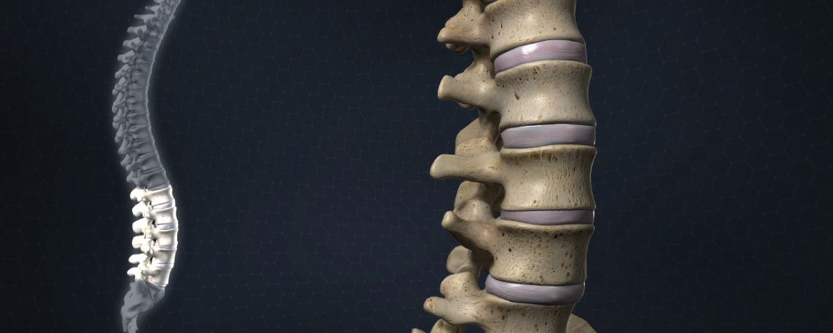
Full spine radiography which is also known as whole spine radiography, is a type of x-ray imaging that evaluates the health of the spine from the neck to the lumbar spine. This type of imaging is typically used to report: abnormal curvature, injuries, tumors, or degenerative diseases.
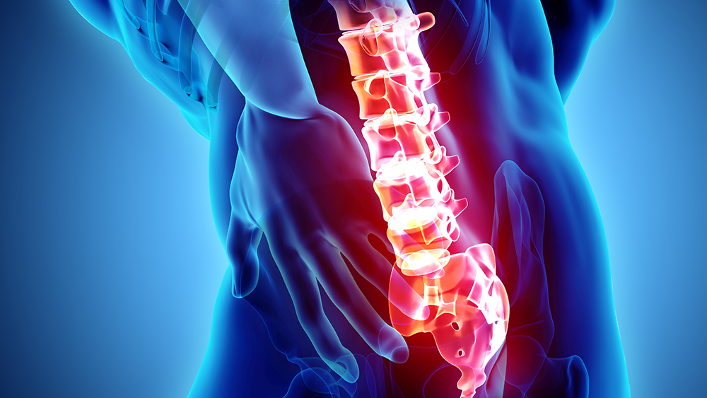
“X-ray imaging of the spine” this is a radiographic medical imaging test using x-rays that provide detailed images (detail views) of neck and lower back bones. People may need an x-ray of the spine if they have structural problems in their spine from birth or have had recent pain from trauma or from diseases such as osteoporosis or arthritis. Spinal radiography may be done for any part of the spine (cervical, thoracic, lumbar, sacral, or coccygeal).
Names of x-ray images of the spine
There are many names for x-ray images of the spine, which depend on which part of the spine is required to undergo imaging; each part of the spine is made up of sections, including:
Cervical spine, made up of seven cervical vertebrae
Thoracic spine, made up of twelve vertebrae in the upper and middle back.
Lumbar spine, made up of five lumbar vertebrae
Sacral vertebrae (sacrum): The sacral bone consists of five small (fused) sacral vertebrae located just above the coccyx.
Coccyx (coccygeal vertebrae): A bone consisting of four coccygeal vertebrae located at the end of the spine.
Based on the patient’s problems, various parts of the spine are imaged (radiographed) as follows; cervical (neck), thoracic (upper back), lumbar (lower back) and sacral (tailbone).
What useful information is obtained from spinal X-ray?
Spinal X-rays provide the doctor with useful information governing many neck and back diseases such as:
Spinal fracture (lumbar fracture).
Osteoporosis (bone miner density).
Arthritis (inflammation of the joint).
Spinal tumor (abnormal cells in the spine).
Slipped disc (if one of the pads between the vertebrae slip out from the vertebrae).
Spinal curvature disorders, which include lordosis (“swayback”), kyphosis (“hunchback”) and scoliosis (when your spine has a side-to-side curvature)
Congenital spinal problems, which include neural tube defects (NTDs) such as spina bifida. Congenital problems are conditions you’re born with.
How X-rays of the spine work
Soft tissues (your skin, organs, and muscles) cannot absorb the X-ray energy that passes through your body. Your bones do absorb the energy. So now your soft tissues are shown as gray in the X-ray image and your bones are shown as white.
Prepping for a spinal X-ray
For the most part, there is nothing special you have to do to prepare for a spinal X-ray. You can eat and drink normal. Your doctor will tell you if you need to do anything special.
It’s a good idea to wear comfortable clothes and leave any jewelry at home. Regardless, you should typically remove and do not keep any jewelry or metal items with you to prevent interference with the X-ray images.
A spinal X-ray usually is done in about 15 minutes. Of course, it might take a little longer depending on how many images need to be taken.
Is the spinal X-ray done standing up?
It depends. For most X-ray images of the spine, you typically will lie on your back on the examination table. This means that while lying on your table the X-ray tube is placed over your body while the X-ray detector (film or detector plate) is placed under your body.
However, in some cases, you may need to stand for a spinal X-ray. The X-ray tube will be positioned in front of you while the X-ray detector is behind you.
Does the X-ray of your spine hurt?
X-rays don’t hurt, but you may feel discomfort during the positioning process if your back has painful or sensitive spots.
Risks and dangers of spinal X-rays
Most people can safely undergo X-ray imaging. High-energy radiation, over time, is damaging to DNA. Occasional x-ray imaging for medical diagnosis, however, is not damaging to DNA because the images are not strong enough.
If you are pregnant, or you think you may be pregnant, you should inform your doctor. Exposure to radiation when pregnant threatens congenital defects in the baby. If the doctor must do an x-ray of the spine, there are specific precautions in place to prevent exposure to the fetus.
Advice after receiving a spine X-ray
Generally speaking, you do not need to do anything special after a spine, back, or neck X-ray. However, your doctor may give additional or different instructions after the X-ray based your specific condition.
