thyroid aspiration biopsy
February 1, 2025
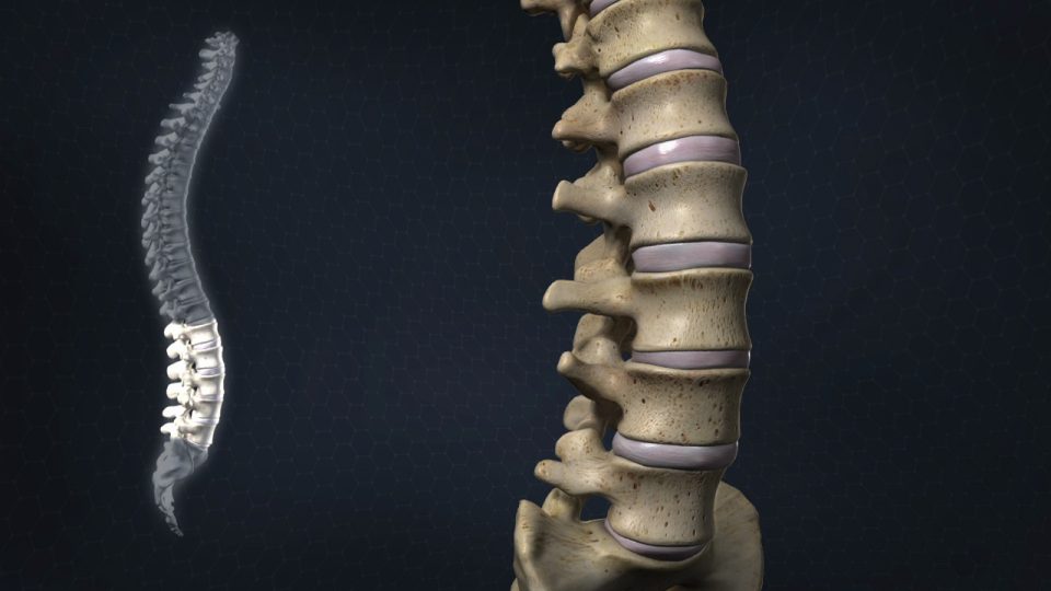
Full spine radiography
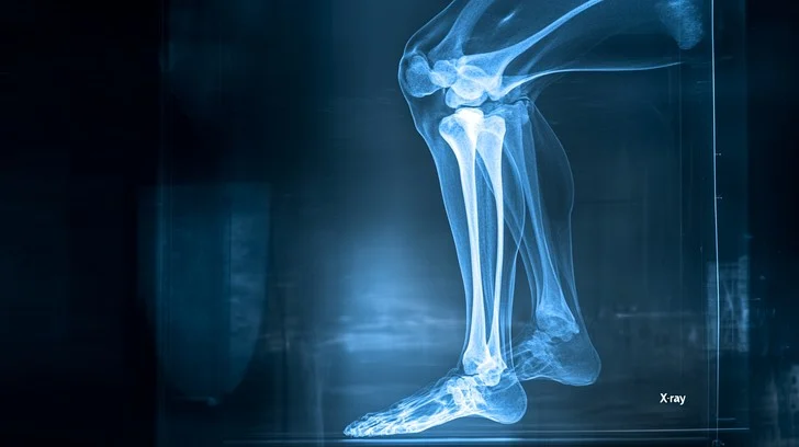
May 18, 2025X-ray imaging of the knee may be a test that makes an picture of the knee’s life structures. Specialists may utilize knee X-rays to analyze and treat conditions of one or both knees. X-ray imaging of the knee could be a speedy, easy, and effortless strategy.
Knee X-ray could be a radiological diagnostic method that employments high-energy beams (X-rays) to supply two-dimensional pictures of the inside structures of the knee (bones).
As specified, knee radiography could be a symptomatic strategy utilized to get pictures of the knee and its bone components such as (tibia, patella) as well as delicate tissues like (muscles, tendons, ligaments, and cartilage) utilizing high-energy X-ray bars. The picture is shaped based on the sum of radiation retained by diverse tissues when uncovered to X-rays.
The bones are exceptionally thick and retain more radiation, showing up nearly totally white.
Delicate and greasy tissues retain less radiation and show up in numerous shades of gray.
Fluids that are radiolucent show up dim in color.

What is an X-ray?
X-rays utilize electromagnetic waves to make an picture of the interior of your body. X-rays are frequently the primary type of imaging utilized when healthcare suppliers are attempting to analyze a illness.
When do I require a knee X-ray?
A healthcare supplier may utilize a knee X-ray to analyze potential wellbeing and therapeutic conditions in one or both knees. A knee X-ray can appear the taking after signs:
Knee bone break (break).
Joint disengagement.
Overabundance liquid that may be a sign of a sprain.
Free bone parts.
Bone goads (osteophyte).
Osteoarthritis.
Irregular arrangement of the knee joint
Bone diseases (osteomyelitis).
Bone diminishing (osteopenia).
Bone cancer.
Moreover, knee X-rays are utilized to screen the recuperating handle after an damage or past surgery.
How could be a knee X-ray performed?
To perform a knee X-ray, the patient’s leg must be situated between the X-ray emitter and the recipient plate (within the conventional and ancient strategy, a radiation-sensitive photographic film is utilized, but nowadays advanced sensors are too accessible). By and large, a few sees of the knee are taken:
Anterior-posterior (AP) or frontal see:
The persistent lies on their back, and the receptor plate beneath the knee and foot is marginally turned internal.
Horizontal see:
The persistent lies on their side with the knee bowed at roughly 30 degrees, and a plate is set underneath it.
Also, depending on the particular case, extra sees may be performed:
Diagonal see:
The quiet lies on their back, the recipient plate is put beneath the knee, and the harmed leg is turned around 45 degrees.
Weight-bearing anteroposterior see:
The quiet stands with their legs straight and parallel, putting their back against the receptor plate.
Pivotal patella see:
The patient’s knee is bowed. This will be done whereas lying on the back with the knee bent, lying down with the bowed leg raised, sitting with bowed knees, or standing with the bowed leg and resting on the examination table over the receptor plate.
It is additionally common to require a photo of the sound foot for comparison and as a reference.
When X-rays are radiated, the receptor plate captures them as an picture. In case an ancient gadget is utilized, a radiology film must be utilized to get the picture. In cutting edge hardware, the picture naturally shows up on the computer in advanced arrange.

What are the perils of X-rays in knee imaging?
X-rays are a speedy and simple way for healthcare suppliers to analyze knee conditions. X-rays contain exceptionally little sums of radiation that pass specifically through your body. Furthermore, X-rays ordinarily don’t cause any side effects.
In case you’re pregnant, your creating child may be exposed to slight radiation. On the off chance that you’re pregnant or arranging to gotten to be pregnant, illuminate your radiologist. You will wear a lead cook’s garment to ensure yourself and your child from radiation. Children are at marginally higher hazard of radiation introduction. Lower measurements may be utilized for children.
Introduction to high levels of radiation carries a slight risk of cancer. In any case, the advantage of precise determination exceeds any chance. In the event that you’re concerned approximately the sum of radiation you may well be uncovered to amid an X-ray, conversation to your technician or healthcare supplier.
Knee radiography could be a totally effortless method. X-rays passing through body tissues don’t cause any distress. Introduction to radiation as it were takes many seconds.
After the examination is completed, the understanding can proceed their work schedule and day by day exercises without any extraordinary care. Typically an outpatient method that does not require hospitalization.
Can an X-ray of the knee show torn knee tendons?
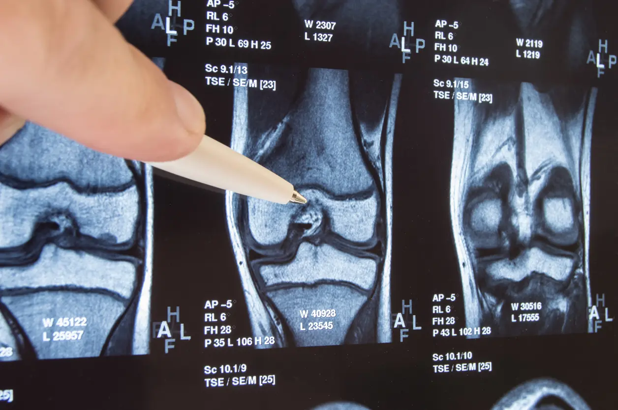
X-rays don’t clearly and precisely appear your soft tissues such as tendons, ligaments, and meniscus. To analyze a tear within the tendons, ligaments, or meniscus, your specialist will ask a computed tomography (CT) filter or magnetic reverberation imaging (MRI). In any case, numerous orthopedic specialists to begin with ask an X-ray to guarantee an exact determination.


