
nuchal translucency (NT) ultrasonography
August 23, 2025
? Is Ultrasound Harmful to the Fetus
February 16, 2026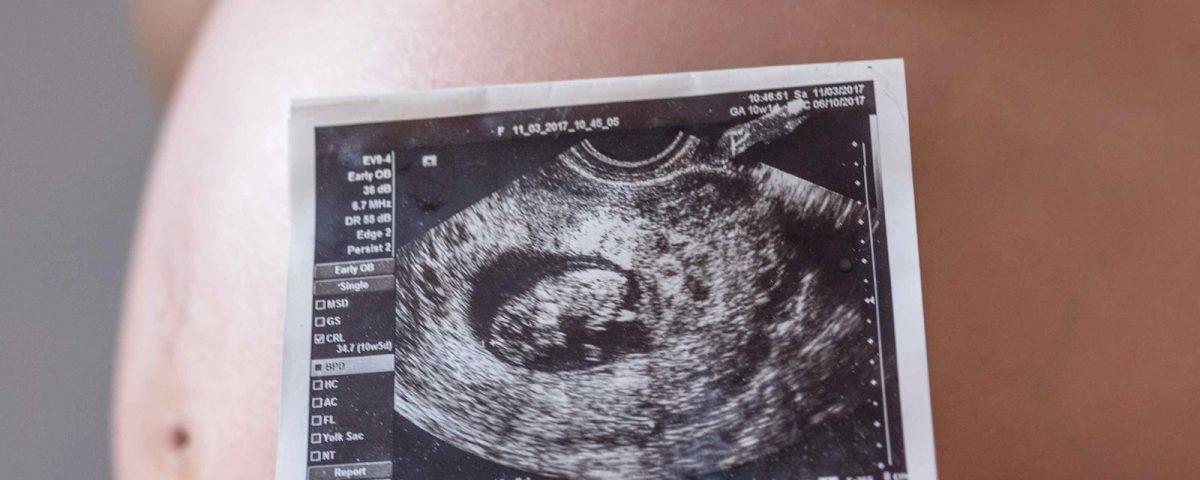
Let’s start exactly where the first concerns of almost every expectant mother begin—at that moment when she wants to be sure everything is progressing normally in her baby’s development. This is where pregnancy ultrasound finds its true meaning: a simple yet incredibly powerful tool that helps us understand how the fetal heartbeat is doing, whether growth is on track, and if the pregnancy is moving forward in a healthy and safe direction. It may look like just a black‑and‑white image, but in reality, it is one of the most reliable methods for monitoring the well‑being of both mother and baby. Ultrasound can ease worries, reveal hidden issues early, and allow for a more precise plan for the rest of the pregnancy.
When a pregnancy moves forward without any imaging or monitoring, certain complications can appear completely silently—from ectopic pregnancy to fetal growth issues, structural anomalies, or conditions that can only be detected in the earliest weeks. This is exactly where the importance of the first‑trimester ultrasound and screening ultrasound becomes clear. These are not just routine checkups; they are the cornerstone of ensuring a healthy pregnancy.
To put it simply, pregnancy ultrasound allows us to see what the naked eye cannot. It opens a small but remarkably accurate window into the developing world inside the uterus. Core keywords in this field—such as pregnancy ultrasound, first‑trimester ultrasound, and screening ultrasound—are rooted in this very stage of care, a type of monitoring that must be both scientifically accurate and emotionally reassuring. It is a form of care every mother deserves.
Introduction to Golestan Imaging Center
If we want to name one of the safest and most trusted places for mothers to have their pregnancy ultrasounds done with complete peace of mind, the Golestan Pasdaran Radiology and Sonography Center is always at the top of the list. For years, this center has specialized in women’s imaging and obstetric ultrasound, and its extensive experience—from the earliest weeks of pregnancy to detailed second‑ and third‑trimester evaluations—has made it one of the most reputable facilities in the Pasdaran area.
One of the key features that sets this center apart is its use of advanced equipment such as the Voluson E10 and the SonoView imaging system. These technologies provide exceptional clarity, fine detail, and the ability to assess even the most delicate fetal structures. Their true value becomes clear when the smallest changes in fetal organs, heartbeat, or growth patterns need to be identified and recorded with precision.
But even the most advanced equipment means little without a skilled team behind it. At Golestan Center, pregnancy ultrasounds are performed under the supervision of Dr. Marjan Zare and Dr. Behnoush Imani, two experienced physicians in women’s health and obstetrics who have spent years refining their expertise in this field. Their precise, attentive approach has earned the trust of countless mothers. The combination of professional expertise, advanced technology, and a calm, supportive environment ensures that each scan is not only accurate, but also reassuring—exactly what every mother needs during pregnancy.
If you’re ready to have your ultrasound done in a professional and trusted setting, you can easily schedule your appointment through the center’s website:
What Is Pregnancy Ultrasound and Why Is It Important?
To put it simply and warmly, a pregnancy ultrasound is the window that lets you see your baby long before birth. But behind this seemingly simple image lies a precise and scientific technology. Ultrasound works through high‑frequency sound waves—completely safe waves that travel into the body through the device’s probe and reflect back from different tissues. The machine then converts these echoes into an image, almost like a quiet yet highly precise conversation between your body and the device.
Unlike X‑ray or CT‑scan imaging, ultrasound uses no harmful radiation. This means you can safely rely on it throughout your entire pregnancy. Another major advantage is real‑time imaging—allowing you to observe fetal movements, the heartbeat, blood flow, and even subtle developmental changes the very moment they happen.
As we step into more advanced forms of ultrasound, 3D and 4D imaging come into play. 3D ultrasound gives a clearer view of the baby’s facial features and structural details, while 4D is essentially 3D with real‑time motion—almost like a short video from inside the womb. Color Doppler also serves an important purpose by showing blood flow in the umbilical cord, fetal heart, and major vessels, helping doctors detect potential circulatory issues early.
Ultrasound plays such a central role in pregnancy care that it’s nearly impossible to imagine the journey without it. From confirming the pregnancy and determining gestational age, to first‑ and second‑trimester screenings, evaluating placental health, monitoring fetal growth, detecting structural anomalies, and assessing amniotic fluid levels—almost everything relies on
ultrasound. The results are always interpreted alongside screening tests, and together they provide a comprehensive, accurate picture of your baby’s well‑being.
Types of First‑Trimester Pregnancy Ultrasounds
The first trimester is always filled with questions, excitement, and those small motherly worries that naturally come with early pregnancy. This is where the very first ultrasounds play an essential role. To get a clear understanding of what’s happening inside the uterus, obstetricians typically rely on two main methods.
Transvaginal ultrasound is the most accurate technique during the earliest weeks. Because the probe is placed closer to the uterus through the vagina, it provides a much clearer image of the implantation site and the formation of the gestational sac. Early vital signs—like the fetal heartbeat—are also usually easier to detect with this method. It is not painful; mothers may only feel a few seconds of mild pressure, which is completely normal.
The second method, transabdominal ultrasound, is the one most mothers recognize. A cool layer of gel is applied to the abdomen, the probe glides gently over the skin, and the developing image appears on the screen. This technique is performed from the outside and is useful for general assessment of the uterus, ovaries, and early fetal growth.
During the first trimester, one of the most important evaluations is determining gestational age through the CRL measurement, which is the crown‑rump length—essentially the baby’s length from head to bottom. This is one of the most accurate indicators of true gestational age, especially up to week 12.
Ultrasound during this period also helps detect critical conditions early, such as ectopic pregnancy or early signs of threatened miscarriage. These early diagnoses can completely change the course of management and relieve significant concerns for the mother.
The ideal time for first‑trimester ultrasounds is usually between weeks 6 and 12. One simple but important point: for a transabdominal scan, it’s better to have a moderately full bladder to improve visibility of the uterus. However, for transvaginal ultrasound, a full bladder is not required.
Key Second‑Trimester Ultrasounds
The second trimester is often the moment when the pregnancy starts to feel real for mothers. They begin sensing fetal movements, physical changes become more noticeable, and ultrasounds become more detailed—and far more exciting. The star of this trimester is the anomaly scan, also known as the detailed anatomy ultrasound. It is arguably the most sensitive and specialized examination of the entire pregnancy.
In this scan, every vital structure is evaluated: the fetal heart with all its chambers and blood flow pathways, the brain and development of its hemispheres, the kidneys and urinary system, long bones, facial structures, the chest, and the spine—which must show a precise and orderly pattern of development.
The anatomy scan is typically performed between weeks 18 and 22—a window when the baby is large enough for detailed visualization but still small enough to be positioned easily for a thorough assessment. This is the best time to detect potential malformations, from cardiac defects to brain anomalies or limb development issues.
This is also when gender determination is usually done. Although the primary goal of the anomaly scan is to assess fetal health and proper organ development, it’s natural for parents to be excited about this sweet detail—and this timeframe offers the most accurate opportunity to determine it.
One of the major advantages of the Golestan Pasdaran Radiology and Sonography Center becomes especially clear during this stage. High‑resolution systems like Voluson provide imaging with exceptional detail—so clear that even non‑experts can notice the difference. The finest fetal structures are visible, allowing diagnoses that are not only precise but also scientifically reliable. This image quality, combined with the extensive experience of the center’s physicians, helps parents leave the anomaly scan with greater peace of mind as they continue their pregnancy journey.
Third‑Trimester Pregnancy Ultrasounds
The third trimester is the final stretch—the stage when mothers need reassurance more than ever, and doctors must clearly understand what conditions are developing inside the uterus. Ultrasounds in this period focus mainly on fetal growth, birth readiness, and placental function.
One of the most important evaluations is EFW (Estimated Fetal Weight), calculated through precise measurements of the head circumference, abdominal circumference, and femur length. These metrics help determine whether the baby is growing normally, growing smaller than expected, or larger than average. This information is critical for planning the timing and mode of delivery.
During this trimester, the position and condition of the placenta are also carefully assessed. Conditions such as placenta previa—where the placenta lies lower than normal—and placenta accreta—where the placenta is abnormally attached to the uterine wall—are identified through ultrasound. Detecting these early helps the medical team plan a safe delivery and minimize risks.
Ultrasound also monitors the amniotic fluid level through the AFI (Amniotic Fluid Index). Too little or too much fluid can indicate specific pregnancy concerns and often guides clinical decisions about further monitoring or intervention.
Another key evaluation in the third trimester is the Biophysical Profile (BPP)—an assessment of fetal movements, practice breathing, muscle tone, and amniotic fluid volume. This test provides reassurance that the baby is developing and thriving in a healthy environment.
For mothers with pregnancy‑induced hypertension or preeclampsia, Doppler assessment of placental circulation becomes especially important. Doppler ultrasound helps evaluate blood flow in the uterine arteries and umbilical cord to determine whether the baby is receiving enough oxygen and nutrients—or if supportive measures are needed.
Doppler Ultrasound in Pregnancy
Whenever detailed evaluation of placental and fetal health is needed, color Doppler ultrasound becomes one of the primary tools physicians rely on. This fascinating imaging technique analyzes blood flow in key fetal and uterine vessels, helping determine whether oxygen and nutrients are reaching the baby properly. The machine sends high‑frequency ultrasound waves and detects their interaction with moving blood cells, converting the flow and direction into color patterns on the screen—essentially turning the dynamics of fetal circulation into a visual story.
During Doppler assessment, several critical vessels are examined. The umbilical artery shows how efficiently oxygen and nutrients are being delivered to the fetus. The middle cerebral artery (MCA) demonstrates how much blood the fetal brain receives, and the uterine arteries reflect how well maternal blood flow supports the placenta. Each vessel serves as a messenger, providing crucial insights into the baby’s overall condition.
One of Doppler’s major roles is diagnosing IUGR (Intrauterine Growth Restriction)—a condition where reduced blood flow limits fetal growth. Doppler is also vital in screening and monitoring mothers at risk of preeclampsia. Increased resistance in uterine artery blood flow can be one of the earliest signs of this condition, making Doppler one of the most reliable methods for early detection.
What sets Doppler ultrasound at Golestan Pasdaran Radiology & Sonography Center apart is the use of Machine‑Based Angle Correction technology. This advanced feature automatically adjusts the angle between the ultrasound beam and the blood flow direction, significantly reducing measurement error. As a result, critical indices such as the S/D ratio and various resistance parameters are reported with exceptional precision. For high‑risk pregnancies or babies requiring close monitoring, this added level of accuracy can meaningfully influence the entire management plan.
۳D and 4D Pregnancy Ultrasound (3D/4D Ultrasound)
If traditional two‑dimensional ultrasound is considered the scientific, diagnostic cornerstone of pregnancy imaging, 3D and 4D scans are its more emotional, visually rich counterparts. The difference lies in how the image is displayed. A 3D ultrasound creates lifelike images of the baby’s face and body with realistic depth and contour, while 4D ultrasound adds real‑time motion—allowing parents to see tiny smiles, yawns, head turns, or even the baby’s fingers gently waving.
Beyond the emotional experience, these technologies hold meaningful diagnostic value. Conditions such as cleft lip, facial anomalies, limb irregularities, and certain surface abnormalities can often be evaluated more clearly with 3D imaging. These images give physicians an additional layer of confidence and help determine whether further evaluation is needed.
For many mothers, seeing their baby’s face before birth is far more than a medical moment—it becomes an unforgettable emotional milestone, one that strengthens the maternal bond and replaces anxiety with joy.
At Golestan Pasdaran Radiology & Sonography Center, 3D and 4D ultrasounds serve both diagnostic and emotional purposes. Advanced Voluson systems produce exceptionally sharp, high‑resolution images—detailed enough to show movements as subtle as fetal blinking. Upon request, these images and live motion clips can be saved as photos or videos, giving families a memory they can cherish forever.
Specialized Ultrasound in High‑Risk Pregnancies
Some pregnancies require closer monitoring from the very beginning or at specific stages—and this is where specialized, high‑precision ultrasound becomes essential. These evaluations are not just about producing clear images; they guide real‑time decision‑making to protect both mother and baby.
One of the most critical concerns in high‑risk pregnancies is placental insufficiency—a condition where the placenta cannot deliver enough oxygen and nutrients. Early identification, especially when Doppler studies of the umbilical artery or uterine arteries show abnormal patterns, allows physicians to plan the safest timing and approach for delivery. These assessments are often combined with serial growth scans to monitor for IUGR (Intrauterine Growth Restriction).
In some pregnancies, especially twin or multiple gestations, fetal growth may become unequal. One fetus may receive less blood flow or have slower development. Such situations require repeated ultrasounds, Doppler studies, and close assessment of amniotic fluid and fetal movements. Multiple pregnancies must also be monitored for complications like TTTS (Twin‑to‑Twin Transfusion Syndrome), making specialized imaging essential.
Pregnancies achieved through IVF also receive tailored ultrasound evaluation. In these cases, physicians not only assess fetal development but also examine implantation quality, placental blood flow, and pelvic structures. IVF pregnancies often require more frequent early‑trimester monitoring to confirm that the gestation is intrauterine and progressing normally.
Another major element in managing high‑risk pregnancies is continuous monitoring of maternal blood pressure and placental circulation. For mothers at risk of preeclampsia, even slight changes in uterine artery Doppler flow can be early warning signs. At Golestan Pasdaran Center, these assessments are performed with high‑resolution equipment and precise Doppler techniques to ensure even the smallest deviations are identified early, allowing timely clinical intervention.
How to Prepare for a Pregnancy Ultrasound
Preparing for a pregnancy ultrasound is much simpler than many mothers expect. A few basic steps can make the entire process smoother and help produce clearer, more accurate images.
For some ultrasounds—especially in early pregnancy—a moderately full bladder can greatly improve visibility by helping ultrasound waves travel more effectively. However, this is not necessary for scans performed after 12 weeks or for transvaginal ultrasounds.
Wearing comfortable, loose‑fitting clothing makes positioning easier and speeds up the imaging process. Many mothers also worry about whether they should stop taking medications before their scan. The important point is: no medication should be stopped without a doctor’s advice. Continuity of treatment is always a priority, and your physician will advise if any adjustments are needed.
Timing also matters. Each scan has its ideal window:
• Dating ultrasound: 6–۱۲ weeks
• Anomaly scan: 18–۲۲ weeks
• Growth scans: typically performed at regular intervals in the third trimester
Understanding these timeframes helps ensure that no critical exam is missed.
From an emotional perspective, going into the ultrasound room with basic knowledge of what will be examined, how long it will take, and what the images mean can significantly reduce stress. At Golestan Pasdaran Radiology & Sonography Center, specialists explain each step calmly and clearly, helping mothers feel at ease and enjoy the moment they see their baby on the screen.
The center follows a structured protocol for pregnant patients, covering preparation, appointment timing, required documents, and supportive guidance to reduce anxiety. From the very first visit, the goal is to provide an experience that feels safe, respectful, and deeply reassuring—while delivering the most accurate diagnostic results possible.

Interpreting Ultrasound Results
When mothers return to receive their ultrasound report, their eyes often land on several English abbreviations and rows of numbers—understandably causing a bit of confusion. But once they know what each term means, reading the report becomes simple and even enjoyable. One of the most common abbreviations is BPD, or biparietal diameter, which measures the width of the baby’s head from one side to the other. It helps evaluate skull development and brain growth.
Right beside it is HC, or head circumference, an essential indicator of fetal brain development and age-appropriate growth. FL, or femur length, is a measurement many parents love seeing because it reflects limb development. AC, or abdominal circumference, is one of the most important parameters in assessing fetal growth and plays a major role in calculating fetal weight.
These measurements are eventually combined in specific formulas to determine the EFW (Estimated Fetal Weight). Physicians compare all these values with the World Health Organization (WHO) growth charts. Normal measurements typically fall between the 10th and 90th percentiles, meaning the baby is growing appropriately compared to others at the same gestational age. When measurements drop below the 10th percentile or show a downward growth trend, doctors consider the possibility of IUGR (Intrauterine Growth Restriction).
This is where the true importance of fetal biometry becomes clear. These numbers are not just measurements—they are the “language” of the fetus. Through these millimeter‑level details, specialists determine whether growth is normal, whether the brain and skull are developing in harmony, whether limbs match gestational age, and whether further evaluation is necessary.
Ultimately, these results gain value only when interpreted alongside the mother’s clinical condition. Radiologists at Golestan Pasdaran Radiology & Sonography Center always take time to explain reports clearly and compassionately, ensuring that mothers understand every part of their pregnancy journey and can plan future follow‑ups with confidence.
How Ultrasound Contributes to Maternal and Fetal Health
Ultrasound is far more than an image—it is a guiding tool in the medical decision‑making process throughout pregnancy. From the earliest weeks, imaging results determine the direction of care: the precise gestational age, the location of placental implantation, the baby’s growth trajectory, and whether everything is progressing as expected. Each image is a vital piece of the puzzle.
During the second‑trimester anomaly scan, for example, detailed evaluations of the fetal heart focus on wall formation, blood flow patterns, and the development of all four chambers. Brain assessment includes the hemispheres, ventricles, and midline structures. For the skeletal system, measurements must match gestational age, and spinal alignment must be perfectly formed—any small deviation matters. The more accurate these details are, the better and more intelligent the medical decisions will be.
Ultrasound plays a major role in increasing the likelihood of a safe delivery. When physicians know that fetal growth is normal, placental function is healthy, amniotic fluid levels are appropriate, and the baby is positioned correctly, the probability of a smooth, low‑risk birth significantly increases. On the other hand, early detection of potential risks like preterm birth, preeclampsia, or structural anomalies allows clinicians to intensify monitoring and provide timely intervention.
Sequential ultrasounds are not only scientifically important but emotionally impactful. Seeing the baby grow, move, and develop heartbeat by heartbeat gives mothers a deep sense of security. This emotional reassurance can transform the pregnancy experience from a stressful journey into a hopeful one.
With a precise yet compassionate approach, Golestan Pasdaran Center ensures that every ultrasound becomes more than a routine exam—it becomes a meaningful moment of connection and informed care for the mother.
Risks, Limitations, and Common Misconceptions About Pregnancy Ultrasound
Ultrasound is one of the safest imaging methods during pregnancy. Contrary to outdated myths, it produces no radiation whatsoever. It works purely through sound waves, similar to everyday environmental sounds but at higher frequencies, which reflect back to form an image. For this reason, global authorities such as the FDA and WHO classify ultrasound as the safest method for fetal evaluation.
Like any medical tool, however, ultrasound does have limitations. In very early pregnancy, for example, the heartbeat may not yet be visible, or fetal movements may be minimal. This is never a sign of a problem—it simply means that more time is needed for the fetus to grow enough to produce a clearer image. This is where the expertise of the physician becomes crucial: knowing when to wait and when additional evaluations are appropriate.
A very common misconception among expectant mothers is the fear of “too many ultrasounds.” Some believe that frequent exams might harm the fetus. In reality, ultrasound produces no cumulative or thermal effects on the body. The energy level of the sound waves is so low that even multiple scans across all trimesters have no impact on fetal development. What truly matters is performing ultrasounds when medically needed, not restricting them unnecessarily.
At Golestan Pasdaran Radiology & Sonography Center, experienced specialists take time to explain these concepts clearly. Many mothers who walk in anxious leave with peace of mind once they understand how ultrasound waves function. Providing this comfort is part of the center’s mission—a commitment not just to imaging accuracy but to guiding mothers through pregnancy with science and compassion.
Periodic ultrasounds are one of the cornerstones of prenatal care. Each scan provides essential insights into fetal growth, placental health, amniotic fluid volume, and organ development. Close monitoring increases the likelihood of a safe delivery and helps mothers experience their pregnancy with greater comfort and confidence.
Over the years, Golestan Pasdaran Center has become one of the most comprehensive prenatal imaging facilities in Tehran. Equipped with advanced systems such as Voluson E10, professional archiving platforms like SonoView, and the expertise of specialists including Dr. Marjan Zarei and Dr. Behnoush Imani, the center delivers high‑precision, reliable results. With a scientific yet compassionate approach, the center provides mothers with a reassuring environment throughout every stage of pregnancy.
For online appointment booking with Dr. Marjan Zarei or Dr. Behnoush Imani, visit:
radiology‑sonography.ir
Or call: 02122562152 _ ۰۲۱۲۲۷۶۵۴۰۷
Frequently Asked Questions (FAQ)
How many ultrasounds are needed during pregnancy?
In most pregnancies, three essential ultrasounds are recommended:
• ۶–۱۲ weeks: to determine gestational age and confirm early viability
• ۱۸–۲۲ weeks: for the detailed anomaly scan
• Third trimester: to assess fetal growth and placental status
In high‑risk or IVF pregnancies, additional scans may be advised depending on the clinical situation.
Does a 4D ultrasound provide greater accuracy?
The main purpose of a 4D ultrasound is to offer real‑time, visually rich images of the baby’s face and movements. While it provides an emotionally meaningful experience, 2D ultrasound and Doppler remain the gold standards for diagnostic accuracy. The 4D scan is considered a complementary tool—not a replacement.
What is the best time to see the baby’s face clearly?
The ideal window is between 27 and 31 weeks. By this stage, a healthy layer of subcutaneous fat has formed, giving the baby’s face its natural contours and allowing for clearer, more lifelike images.
Do all insurance companies cover pregnancy ultrasounds?
Many insurance providers do cover a significant portion of essential pregnancy ultrasounds. Coverage varies based on the specific insurance plan and the type of ultrasound needed. At Golestan Pasdaran Center, mothers receive full guidance about coverage before their appointment.
Can all pregnancy ultrasounds be performed at one specialized center?
Yes—and in fact, this is the best option. Completing all pregnancy scans in a single specialized center helps maintain a consistent imaging record and significantly improves the accuracy of growth trend evaluations. Golestan Pasdaran Radiology & Sonography Center is one of the few facilities equipped to perform all types of pregnancy imaging—from screening ultrasounds to Doppler and 4D scans—using advanced technology and a highly trained team.