
thyroid ultrasound
May 18, 2025
Guide to Pregnancy Ultrasound Types and Their Clinical Applications
November 21, 2025
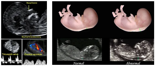
During the first trimester of pregnancy, the nuchal translucency (NT) ultrasonography is an elective procedure. This test aids in assessing the likelihood that the fetus will have congenital conditions like Down syndrome. This test has a high diagnostic accuracy when used in conjunction with other first-trimester tests. Late in the first trimester and early in the second trimester (between 11 weeks and 3 days and 13 weeks and 6 days), a subcutaneous space filled with natural fluid is seen at the back of the fetus’s neck. This condition is known as nuchal translucency.
Nuchal translucency: what is it?
An ultrasound technique called nuchal translucency (NT) gauges the volume of fluid behind the fetus’s neck during the first trimester of pregnancy. Measuring the amount of fluid can assist in determining the probability of a chromosomal or genetic disorder in the fetus. A tiny amount of fluid is normal. NT ultrasonography is not a disease diagnosis; rather, it is a screening test. It assists your doctor in determining whether additional testing is necessary and whether the fetus is at risk. The nuchal fold, which is located behind the fetus’s neck, is examined via the nuchal translucency scan. There is fluid at the back of every fetus’s neck.
Healthcare providers have discovered that increased fluid accumulates at the neck’s base of a fetus with chromosomal or genetic disorders. Increased fluid in this region may suggest that the fetus has disorders like Down syndrome (trisomy 21), Patau syndrome (trisomy 13), or Edwards syndrome (trisomy 18). An elevated NT measurement might also suggest a greater likelihood of congenital heart conditions. The outcomes of the NT scan can indicate if the fetus is at risk for these diseases. During nuchal translucency, the ultrasound additionally examines several essential anatomical features of the developing fetus. Additional anomalies detected during NT could elevate the risk of genetic or structural disorders.
Pathology
It is believed that the increase in nuchal translucency is associated with the dilation of lymphatic vessels and is considered a non-specific sign of a more general fetal anomaly. Measuring nuchal translucency requires specific and standardized evaluation and careful attention to techniques. The connection of this issue with diseases The nuchal translucency measurement can be associated with a number of abnormalities, including:
Aneuploidy
Trisomies (including Down syndrome)
Turner syndrome
Non-aneuploid structural defects and syndromes
Congenital heart disease
Noonan syndrome: the only genetic molecular disorder with a clear association with increased nuchal translucency.
Congenital diaphragmatic hernia
Omphalocele
Skeletal dysplasias
Smith-Lemli-Opitz syndrome
Miscarriage or fetal death
Intrauterine infections
When is the NT ultrasound conducted?
The NT scan is conducted by the doctor during weeks 11 to 13 of pregnancy, or when the fetus measures between 45 to 84 millimeters from crown to rump (lower body). The explanation for this is that the fluid located behind the fetus’s neck is usually reabsorbed by the body following the 14th week of pregnancy. This complicates its measurement in the later stages of pregnancy. NT tests are frequently conducted as a component of first-trimester screening assessments.
How to conduct the NT ultrasound
The healthcare provider employs an abdominal ultrasound (or occasionally a vaginal ultrasound) to measure the nuchal translucency. Initially, they apply ultrasound gel to your stomach. Next, they glide a transducer (a hand-held device) across your abdomen. Pictures of the fetus are shown on the screen. Measurements of the fluid-filled area located behind the neck of the fetus are recorded. The fluid’s area is measured in millimeters.
The outcomes of the NT scan are typically not assessed in isolation. The physician typically merges the results of all your initial-trimester tests to determine the total risk of the fetus having a birth defect. Typically, a blood test in conjunction with the NT scan is advised to enhance the screening’s accuracy. If the blood test is not conducted, your age along with the fetal nasal bone may also be risk factors.
It is important to note that the physician does not identify the illness solely from the NT scan. NT scans serve as screening tests solely to assess the probability of possessing a disease. If NT is high, your physician or genetic counselor may explore further testing possibilities, such as chorionic villus sampling (CVS). Often, a higher NT does not indicate a chromosomal or genetic issue, so further testing is generally advised.
What is the accuracy of the nuchal translucency test?
NT screenings by themselves can identify roughly 70% of instances of trisomy 21 or Down syndrome. Numerous healthcare providers integrate the conventional NT ultrasound with blood tests. When first-trimester blood tests are combined with the accuracy of predicting conditions, the result rises to approximately 95%.
Are there any risks associated with NT ultrasound?
No, the nuchal translucency ultrasound poses minimal risk as a screening method.
Additional assessments
If the findings are unusual, additional examinations involve the following:
Amniocentesis or chorionic villus sampling.
Fetal heart ultrasound
What does an unusual NT signify?
NT screenings solely reflect the likelihood of the fetus acquiring a condition. If your screening shows abnormalities, your doctor might employ a test like chorionic villus sampling (CVS) or amniocentesis (during later pregnancy) to identify the disease.
No need to be concerned. An abnormal result does not necessarily indicate an issue with the fetus. Your physician will perform additional tests or search for other indications of issues in your ultrasound or blood examination.
What is the standard measurement for the fetal nuchal translucency?
This indicates that the fetus faces increased risk or a higher chance of developing these conditions.