Elastography
November 24, 2024
thyroid aspiration biopsy
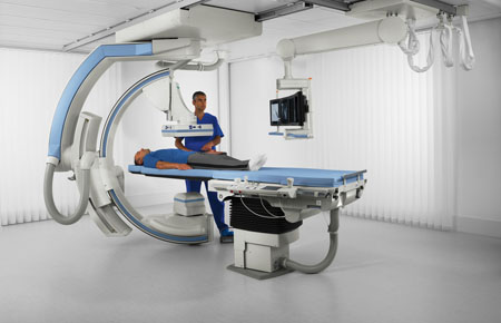
February 1, 2025Fluoroscopy is a type of X-ray imaging method that shows the movement of organs, tissues, or other internal structures in real-time. The fluoroscopy method is like a video. Fluoroscopy allows doctors to observe the functioning of organs or structures such as heart rate, lung function, or bowel movement while food is in motion. This method can help your doctor evaluate various conditions and diagnose diseases.
Doctors can use fluoroscopy to observe several body systems in real-time, including:
Cardiovascular system.
Digestive system.
Urinary system.
Musculoskeletal system.
Reproductive systems.
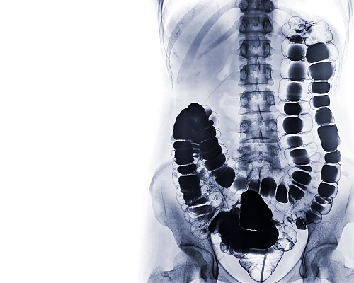
Fluoroscopy applications
Doctors use fluoroscopy for diagnostic purposes and visual guidance during certain procedures (as interventional guidance).
Diagnostic fluoroscopy:
Doctors use fluoroscopy for various parts of your body to diagnose several diseases, including:
Barium swallow (barium swallow and barium meal): A barium swallow is a fluoroscopic imaging test that examines problems in the upper gastrointestinal (GI) tract, which includes the mouth, back of the throat, esophagus, stomach, and the first part of the small intestine. This test involves drinking a chalky-tasting liquid that contains barium, a safe substance that makes parts of your body show up more clearly on X-ray imaging. These tests can help diagnose esophageal disorders, ulcers, hiatal hernias, GERD (gastroesophageal reflux disease), structural problems in the digestive tract, and tumors.
Barium enema: A barium enema, also known as a lower gastrointestinal (GI) series, is a fluoroscopic imaging test that examines problems in the colon and rectum (parts of the large intestine). A safe liquid containing barium is introduced into the body through a tube inserted into the rectum. This liquid coats the inside of the colon and clearly outlines its shape in X-ray imaging. This test can help diagnose inflammatory bowel disease (Crohn’s disease or ulcerative colitis), diverticulosis, colon cancer, polyps, and volvulus of the colon (abnormal twisting of the intestine).
Angiography: Angiography or angio uses fluoroscopy to identify and diagnose narrowing or blockage of blood vessels in the body.
Cystography: Cystography is an imaging test that can use fluoroscopy to help diagnose bladder problems. During a cystography, the healthcare provider inserts a thin tube called a urinary catheter into the urethra and injects contrast dye into the bladder. This dye helps certain parts of the bladder to appear clearer in X-ray imaging. This test can help the doctor examine bladder emptying during urination. This is also called a voiding cystogram.
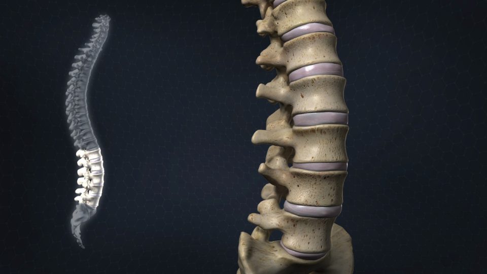
Myelography: Myelography uses fluoroscopy and the injection of contrast material to evaluate the spinal cord, nerve roots, and the spinal covering (meninges). This is especially useful for evaluating the spine after surgery and for assessing disc problems in individuals who cannot undergo an MRI test.
Hysterosalpingogram: In this procedure, a provider uses fluoroscopy to provide images of the female reproductive organs. It can help diagnose some causes of infertility.
Fluoroscopy as a visual guide
Cardiac catheterization: In this method, fluoroscopy shows blood flow in the arteries. It can help visually guide doctors in performing angioplasty.
Placement and guidance of catheters: Fluoroscopy can help ensure the proper placement of catheters, which are thin, hollow tubes, to allow fluids to enter or exit your body. Providers can place catheters through the urinary tract, blood vessels, and bile ducts.
Stent placement: Fluoroscopy can help ensure the proper placement of stents, which are devices that help open narrowed or blocked blood vessels.
Orthopedic surgery: The surgeon may use fluoroscopy to help guide orthopedic procedures such as joint replacement and fracture repair (broken bone).
How fluoroscopy is performed
Depending on the type of procedure, fluoroscopy may be performed at an outpatient radiology center or as part of your hospital stay. This procedure may
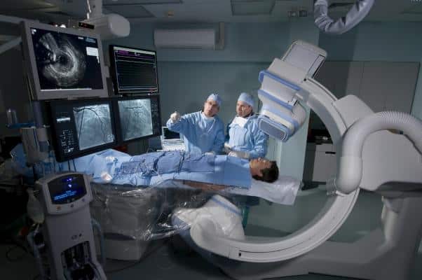
involve some or most of the following steps:
You may need to remove any jewelry, removable dental appliances, glasses, and metallic objects that might interfere with this procedure.
You may need to remove your clothing. If so, a hospital gown will be given to you.
Depending on the type of fluoroscopy, you may be given a lead shield or apron to wear over the pelvic area or another part of your body.The apron protects against unnecessary radiation.
For some procedures, you may be asked to drink a liquid containing contrast dye. Contrast dye is a substance that makes parts of your body show up more clearly on an X-ray.
If you are not asked to drink a colored liquid, the dye may be injected through an intravenous (IV) line or introduced into the body through an enema.
You will be placed on a table. Depending on the type of procedure, you may be asked to move your body into different positions or move a specific part of your body. You may also be asked to hold your breath for a short period of time.
If your procedure involves taking a catheter, your provider will insert a needle into the appropriate part of your body. This site may be the groin, elbow, or another location.
Your provider uses a specialized X-ray scanner to create fluoroscopic images. Specialists can view them on a computer screen.
If a catheter has been placed, the specialist will remove it.
In some procedures, such as those involving injection into a joint or artery, a painkiller or a sedative may be administered first. For procedures that use fluoroscopy as an imaging guide during surgery or stent placement, general anesthesia may also be used to calm you.
What are the risks of fluoroscopy?
Fluoroscopy has some of the same risks as other X-ray methods due to radiation exposure. For this reason, if you are pregnant or think you might be pregnant, you should not undergo a fluoroscopy procedure. Radiation can be harmful to a developing fetus.
If used appropriately, fluoroscopy for diagnostic purposes results in very low levels of radiation exposure.
When specialists use fluoroscopy for certain invasive or surgical procedures, it may result in higher levels of radiation exposure. The radiation-related risks associated with fluoroscopy for these purposes include:
Radiation injuries to your skin and underlying tissues (“burns”), which occur shortly after exposure to radiation.
Radiation-induced cancers, which may occur later in life.
The likelihood of experiencing these side effects is very low. If this procedure is medically necessary, the benefits of the procedure outweigh the potential risks of radiation.
If the contrast dye is part of your fluoroscopy procedure, there is a small risk of an allergic reaction. If you have an allergy or have ever reacted to contrast materials, be sure to inform your doctor.
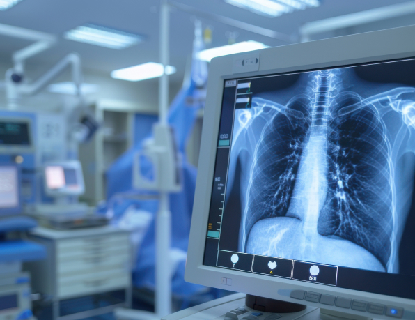
Interpretation of fluoroscopy results
Your results depend on the type of procedure you have undergone. Several diseases and disorders can be diagnosed with fluoroscopy. The doctor may need to send your results to a specialist or conduct further tests to aid in the diagnosis.
If you have questions about your results, talk to your doctor. To understand the results of the fluoroscopy method, the doctor may also consider symptoms, medical history, and the results of other tests.

