
liver ultrasound
November 24, 2024
Hysterosalpingography
February 1, 2025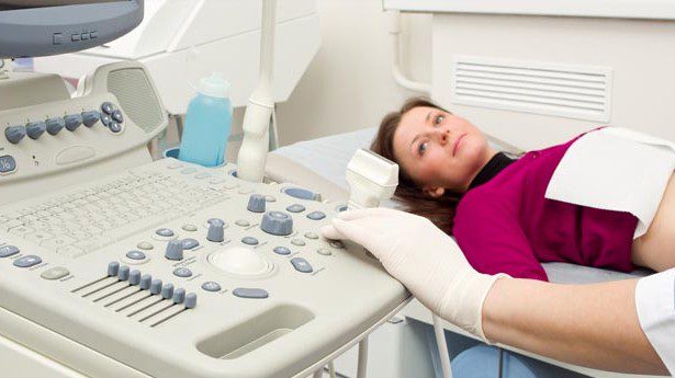
Infertility, recurrent miscarriages, and unexplained vaginal bleeding are just a few of the conditions that can be diagnosed by hysterosonography, sometimes referred to as sonohysterography, which employs sound waves to make images of the inside of the uterus. A standard examination of a woman is remarkably similar to hysterosonography.
Another name for sonohysterography is saline infusion sonography. One possible component of the sonohysterography evaluation is Doppler ultrasound. A unique ultrasound technique called Doppler ultrasound measures blood flow as it moves through a blood artery.
A sonohysterogram: what is It?
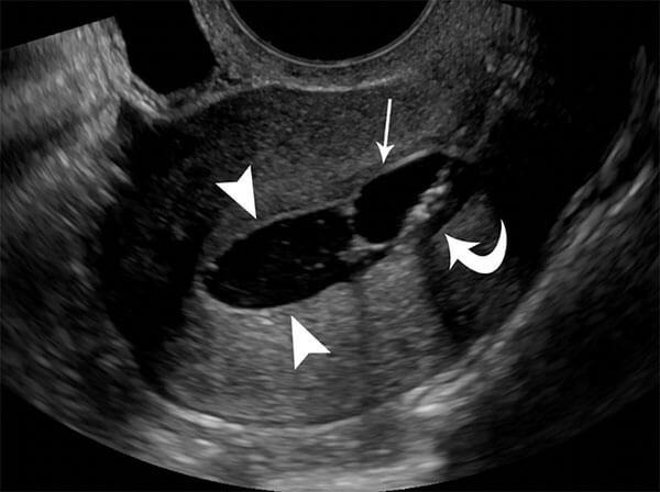
A particular kind of ultrasound that gives the doctor a view inside the uterus is called a sonohysterogram. While the uterus is filled with saline, a sonohysterogram uses ultrasound to record the shapes and structures inside the uterus. Your doctor can use a sonohysterogram to view trouble spots in the uterus and the endometrium, the uterine lining, which might result in unintended symptoms including infertility, pelvic pain, and bleeding. Another frequent name for this process is SIS.
Some common applications of the hysterosonography method
This is a valuable technique for evaluating unexplained vaginal bleeding, which may be the result of uterine abnormalities such as the following:
Polyp
Fibroids
Endometrial adhesions (or scarring)
Lesions / malignant masses
Congenital defects
Sonohysterography is also used to examine uterine abnormalities in patients who experience infertility or multiple miscarriages.
Doppler ultrasound images can also help the doctor evaluate the following:
Blood flow obstruction (such as a clot).
Blood flow in polyps, tumors, and abnormalities.
Pelvic varicose veins and aneurysm.
The best time to perform a sonohysterogram
Sonohysterograms are ideally performed between days 6 and 11 of the cycle, after the menstrual period ends but before ovulation. The doctor can see the photos clearly since the uterine lining is at its thinnest at this time. It can also be done at any moment if you are going through menopause.
For whom is a sonohysterogram not appropriate?
People with pelvic inflammatory disease (PID) and those who are pregnant shouldn’t have a sonohysterogram.
Tips for getting ready for a hysterosonography
Dress comfortably and loosely.
For this process, you might need to wear specific attire. No specific preparation is required before the test.
To reduce any possible discomfort, you could be told to take an over-the-counter medicine just before the surgery.
The physician inserts a catheter into the uterus and a speculum into your vagina. The doctor uses an ultrasound transducer to inspect the uterine lining and injects a small amount of sterile saline into the uterine cavity through a tiny tube that is placed into the vagina. Soft tissues that are difficult to see on X-rays can be clearly seen using ultrasound, which has no known negative effects.
With the exception of taking pictures as a tiny catheter gently inserts a few tablespoons of sterile saline into the uterus, the sonohysterogram technique is somewhat similar to the transvaginal ultrasound technique. A transducer is inserted into the cervix and vagina, just like in a transvaginal ultrasound. Once inside, it releases sound waves that record details about the uterine structures. The radiologist doing your test can then view an image of the uterus’s appearance on a screen using this information.
What dangers might a sonohysterogram pose?
There are few adverse effects and a low risk associated with a sonohysterogram. Taking painkillers in advance will assist ease any discomfort you may have during specific parts of the treatment. Although there is a slight risk of infection, your physician can take precautions.
Keep an eye out for these warning indicators that could point to an infection:
Damn.
Severe pain.
Changes in vaginal discharge
If you notice these symptoms, inform your doctor immediately.
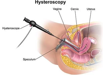
What distinguishes a hysteroscopy from a sonohysterogram?
The doctor can view inside the uterus using both a sonohysterogram and a hysteroscopy. Compared to a hysteroscopy, a sonohysterogram is faster, less intrusive, and less expensive. It is solely useful for diagnostic purposes, though. Through hysteroscopy, the physician can view the inside uterine structures and, if required, remove them all at once.
What distinguishes a hysterosalpingography (HSG) from a sonohysterogram?
Hysterosalpingography (HSG) visualizes and displays the uterine structures on a screen using X-ray radiation rather than sound waves. HSG can reveal whether your infertility issues are being caused by a blockage in the fallopian tubes. A sonohysterogram may not always show these obstructions.