Doppler ultrasound
November 24, 2024
sonohysterography
February 1, 2025A liver ultrasound can provide a lot of information about the liver. Doctors may order a liver ultrasound to screen for liver diseases or to assess how well a treatment is working. Sometimes, an ultrasound alone is sufficient to diagnose or rule out these conditions, but at other times, it may lead to further tests.
Liver ultrasound is a non-invasive test that uses ultrasound waves sent towards the internal organs to create images of the liver and blood vessels. Liver ultrasound can help diagnose various diseases affecting the liver, such as fatty liver and gallstones.
Liver ultrasound is safe and usually performed quickly, taking up little time. However, some people may feel anxious about undergoing a liver ultrasound due to concerns about what the test might reveal.
Reasons for performing a liver ultrasound
If you have symptoms of liver disease, such as abdominal pain, abnormal enlargement and swelling of the abdomen, or if other test results indicate liver disease (such as a blood test), your doctor may recommend a liver ultrasound. Ultrasound provides a quick and easy way for doctors to examine these findings. If you have already been diagnosed with liver disease, your doctor may order a liver ultrasound to check the condition of your liver.

Can an ultrasound show liver problems?
A liver ultrasound can show signs of fat accumulation in the liver (steatosis), inflammation and swelling (hepatitis), and scar tissue (fibrosis or cirrhosis). These three are the main chronic liver diseases. Ultrasound may also show liver lesions and abnormal spots on the liver. Certain types of liver ultrasound can also assess blood flow in the liver or the stiffness of its tissues.
Liver ultrasound can be useful in diagnosing many liver diseases:
Chronic liver diseases:
Hepatitis
Steatosis
Cirrhosis
Liver lesions:
Liver cysts
Liver hemangioma
Liver cancer
Liver vascular conditions:
Ischemia
Budd-Chiari syndrome (the hepatic vein is narrowed or blocked by a clot)
Portal hypertension
Other conditions that affect your biliary system:
Ascites (fluid accumulation in the abdominal cavity)
Cholecystitis (sudden inflammation of the gallbladder)
Cholecystolithiasis (gallstones)

Preparation for a liver scan
Liver ultrasound does not require any special preparation. Since it is a low-risk and non-invasive method, the individual does not need painkillers or anesthesia. However, the person may be asked to fast before the ultrasound, especially for the gallbladder and liver, because this can cause the gallbladder to swell and make it easier to see.
Different types of liver ultrasound
Doctors may combine liver ultrasound with other techniques to obtain a more accurate image and diagnose potential problems. Some common types of liver ultrasound include:
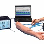
Doppler imaging: Doppler ultrasound is a non-invasive method (without needles and injections) used to estimate blood flow from blood vessels. This method can be particularly useful for detecting growths and lesions on the liver and diagnosing liver cancer.
Elastography: It is a technique for observing the stiffness of liver tissue, which can be a sign of cirrhosis or other problems.
Combined techniques: The doctor may combine techniques such as performing an ultrasound and an MRI scan.
How to perform a liver ultrasound
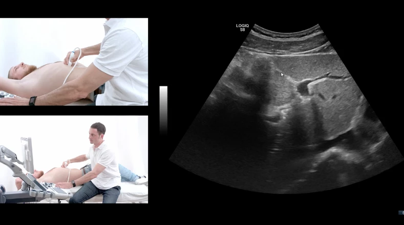
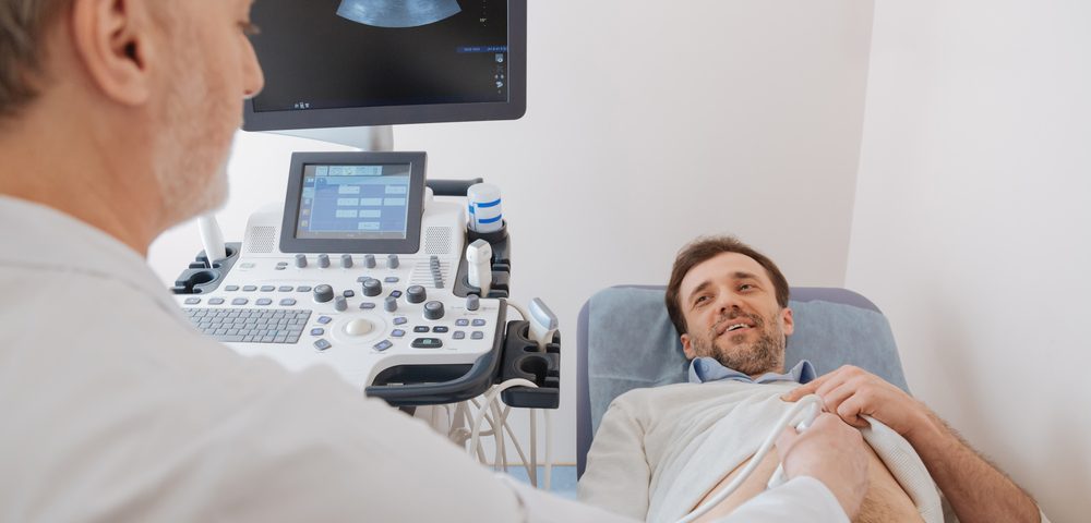
A liver ultrasound requires access to the person’s abdomen. You may wear a hospital gown or just lift your shirt. During the ultrasound, the person lies on a raised table, and the ultrasound technician rubs a transducer on the abdomen. The transducer is a small handheld device that resembles a mace. The transducer sends images to the computer. The person themselves might also be able to see the images simultaneously. The ultrasound specialist might also examine the gallbladder during the scan. Both organs are located in the upper right part of the abdomen and near the ribs.
Some people may feel pressure during the test or feel anxious about the results. However, a liver ultrasound will not harm you. Cold gel may be used, which can be somewhat uncomfortable.
Ultrasound usually only takes a few minutes.
After a liver ultrasound, there is no need for recovery time. The person can eat, drink, drive, and return to work or home after the ultrasound.
Does a liver ultrasound have any risks or side effects?
Liver ultrasound has no side effects. The only issue could be that it is not done correctly because it is a method that also depends on the expertise, skill, and experience of the person performing it. Since a liver ultrasound is usually the first test your doctor uses to screen for liver problems, you don’t need to worry too much about the small risk of inaccurate results or insufficient accuracy in performing it.
Interpretation of liver scan results
Results of a normal versus abnormal liver ultrasound
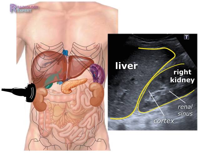
A normal liver in an ultrasound has a specific expected size, shape, and color (gray shadow). The radiologist can measure the size and shadow with other adjacent organs to compare them. The surface should be smooth and soft, not lumpy or coarse. Lumps or spots on the surface of the liver may indicate cysts or solid masses. The radiologist may also look for enlarged blood vessels or bile ducts.
Enlargement or shrinkage of the liver indicates liver disease. The mass surface indicates scarring (cirrhosis) or a tumor. Some lesions are natural, but they shouldn’t be too many. The brightness or darkness of the liver tissue color is also important; for example, fatty liver disease may make it lighter, while inflammation may make it denser and darker. Your ultrasound report explains what your radiologist sees.


