tests and examinations related to pregnancy
October 8, 2024
Fluoroscopy
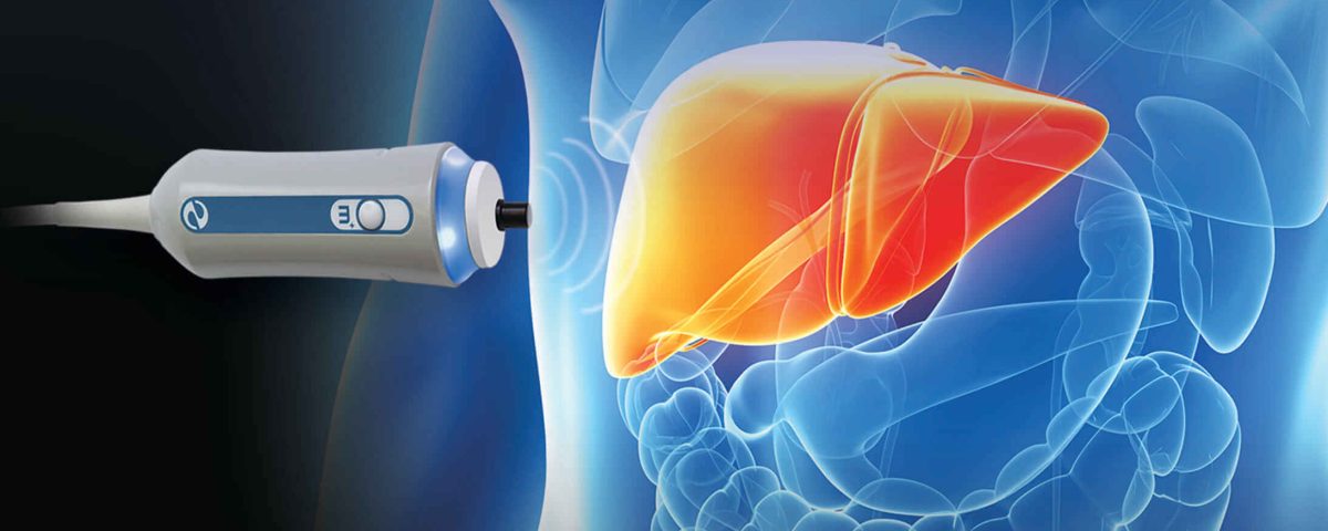
November 24, 2024Elastography is a test used to examine the “elasticity” of body organs, especially the liver. This test helps doctors accurately diagnose and stage specific conditions such as liver fibrosis. Elastography is a type of imaging test that examines organs to determine if they are stiffer than normal. Stiff areas in your organs may be a sign of disease. Elastography is primarily used to assess liver stiffness. Stiff areas in the liver are a sign of scar tissue (fibrosis) caused by liver disease.
Liver fibrosis occurs when certain diseases gradually damage the liver over time. When the liver tries to heal, it forms more scar tissue. Many conditions that cause ongoing or repeated liver damage can lead to liver fibrosis.
Conditions that usually cause fibrotic disease include:
Hepatitis C and Hepatitis B
Alcohol Use Disorder (AUD)
Non-alcoholic fatty liver disease (NAFLD)
With the formation of fibrosis, scar tissue replaces healthy liver cells and prevents the normal functioning of the liver. Scar tissue can reduce blood flow in the liver. Lack of blood flow causes further liver damage and increased fibrosis.
Without treatment, fibrosis can become severe. Severe fibrosis is called cirrhosis. With cirrhosis, liver scarring is permanent and can lead to serious health issues, including liver failure. Cirrhosis also increases the risk of developing liver cancer.
Fibrosis and early cirrhosis do not always cause symptoms. But elastography can help detect liver scarring early, so treatment can begin before severe damage occurs.
There are two types of liver elastography tests:
Ultrasound elastography: which is also called transient elastography. An ultrasound device uses sound waves to send vibrations to your liver. This device measures the speed of vibrations moving through your liver. If there are areas of stiffness in the liver tissue, the vibrations in that area move faster. A computer uses the measurements to create an image that shows any stiffness in your liver tissue, which is a sign of fibrosis.
MRE (Magnetic Resonance Elastography): Sends vibrations to your liver that are measured with Magnetic Resonance Imaging (MRI). MRI is a method that uses strong magnets and radio waves to create images of the organs and structures inside the body. In the MRE test, a computer program creates a map showing each stiff area of your liver.
Other names for these tests include: liver elastography, transient elastography, FibroScan, MR elastography.
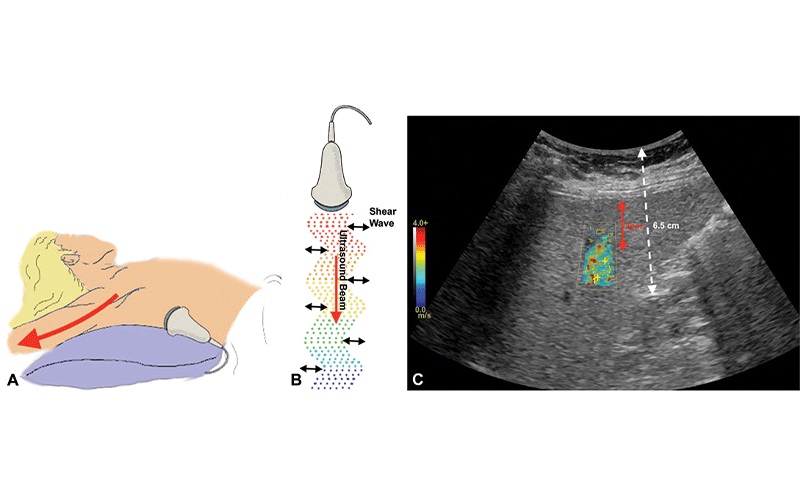
What is shear wave elastography?
Shear wave elastography is a newer ultrasound technology used to assess tissue stiffness. This method produces higher quality images. This method is still under development; shear wave elastography is used in ultrasound for the following:
Chest
Liver
Musculoskeletal system
Prostate
Thyroid nodule
What is the purpose of elastography?
The most common reason for elastography is to check the liver for fibrosis, which occurs in the early stages and may progress to the final stage or liver cirrhosis. If left untreated, liver fibrosis can lead to serious health problems such as:
Cirrhosis
Liver cancer
Liver failure
Gastrointestinal bleeding
It happens.
How accurate is liver elastography?
While research varies, most specialists agree that elastography can accurately diagnose liver fibrosis. Many doctors use elastography to diagnose this disease. More importantly, it is also used to monitor the progression of the disease over time.
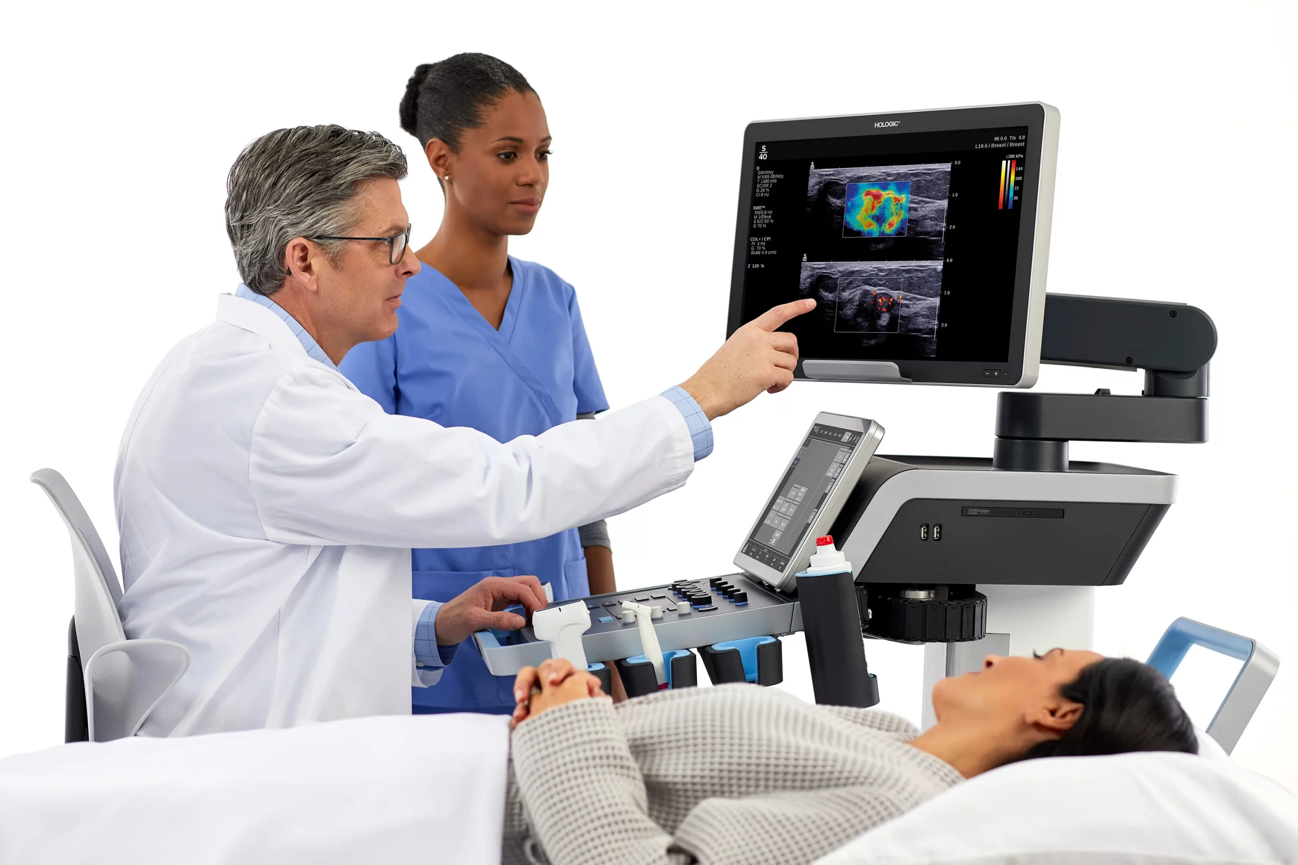
How to perform elastography
Elastography ultrasound:
The elastography test is quick and usually involves the following steps:
Removing clothing from the abdomen to expose the skin over the liver area.
You lie on a table, and the doctor spreads a special gel on your skin.
While the device sends a series of vibrations to your liver, you will hold your breath for 10 to 15 seconds.
The vibrations pass through your liver and return to the device that sends the information to the computer.
The computer creates an Image of your liver that shows any areas of stiff tissue and other signs of liver disease.
MRE (Magnetic Resonance Elastography): performed with an MRI machine. During the MRE procedure:
You lie on a narrow table.
The technician places a few small devices on the lower right side of you’re abdomen. A device called a “driver” sends painless vibrations to your liver. Other devices send and receive radio waves to and from the computer.
The table slides into the MRI machine, which is like a large tunnel. When the machine takes pictures, you will hear loud sounds. You may be given earplugs or headphones to reduce the noise. Inside the MRI machine, you will be asked to hold your breath for 10 to 15 seconds while the vibrations pass through your liver. The computer creates images of your liver that show any scars or other signs of liver disease.
Risks of elastography
There are no known risks associated with elastography ultrasound. When appropriate safety guidelines are followed, MRE also poses no risk to the average patient. The magnetic field is not harmful. If you are receiving a sedative, there are risks associated with excessive sedation. There is also a small risk of an allergic reaction to the gadolinium contrast solution, which is only used for certain MRI procedures.
Interpretation of elastography results
Elastography produces an image of your liver. The Image shows the stiffness level to the radiologist, indicating the presence of scarring. The scar level varies from mild to advanced and includes the following:
F0 to F1: no scars or mild scars
F2: Moderate scar
F3: Severe scarring
F4: Advanced scarring (cirrhosis)
The results also indicate how much fat has accumulated In the liver. The measurement of liver fat is called the “CAP score.” The”amount of fat in the liver under normal conditions Is 5 percent or less. Higher amounts indicate the presence of fatty liver disease, which may lead to fibrosis. If you have any questions about your test results, talk to your doctor.

