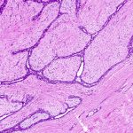
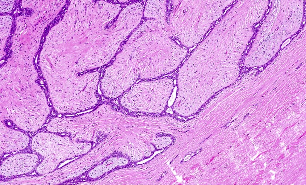
Breast fibroadenoma from diagnosis to treatment
June 28, 2025
Fatty liver disease is one of the most common conditions of our time—a disease that progresses silently, usually without pain, and precisely for that reason is often underestimated. Statistics show that more than 30% of Iran’s adult population has some degree of fatty liver disease; an alarming figure that has been rising in recent years due to obesity, diabetes, and sedentary lifestyles. But the key question is this: Are all fatty livers harmless? Or do some of them quietly progress toward more serious damage?
Many people, after undergoing an ultrasound examination, are told only one sentence: “You have grade 1 or grade 2 fatty liver.” This statement often creates a false sense of reassurance—as if the condition is minor and not worth worrying about at the moment. However, the reality is that fatty liver disease is not always what appears on ultrasound. The more important issue is what cannot be seen easily: the degree of liver stiffness and the possible onset of liver fibrosis.
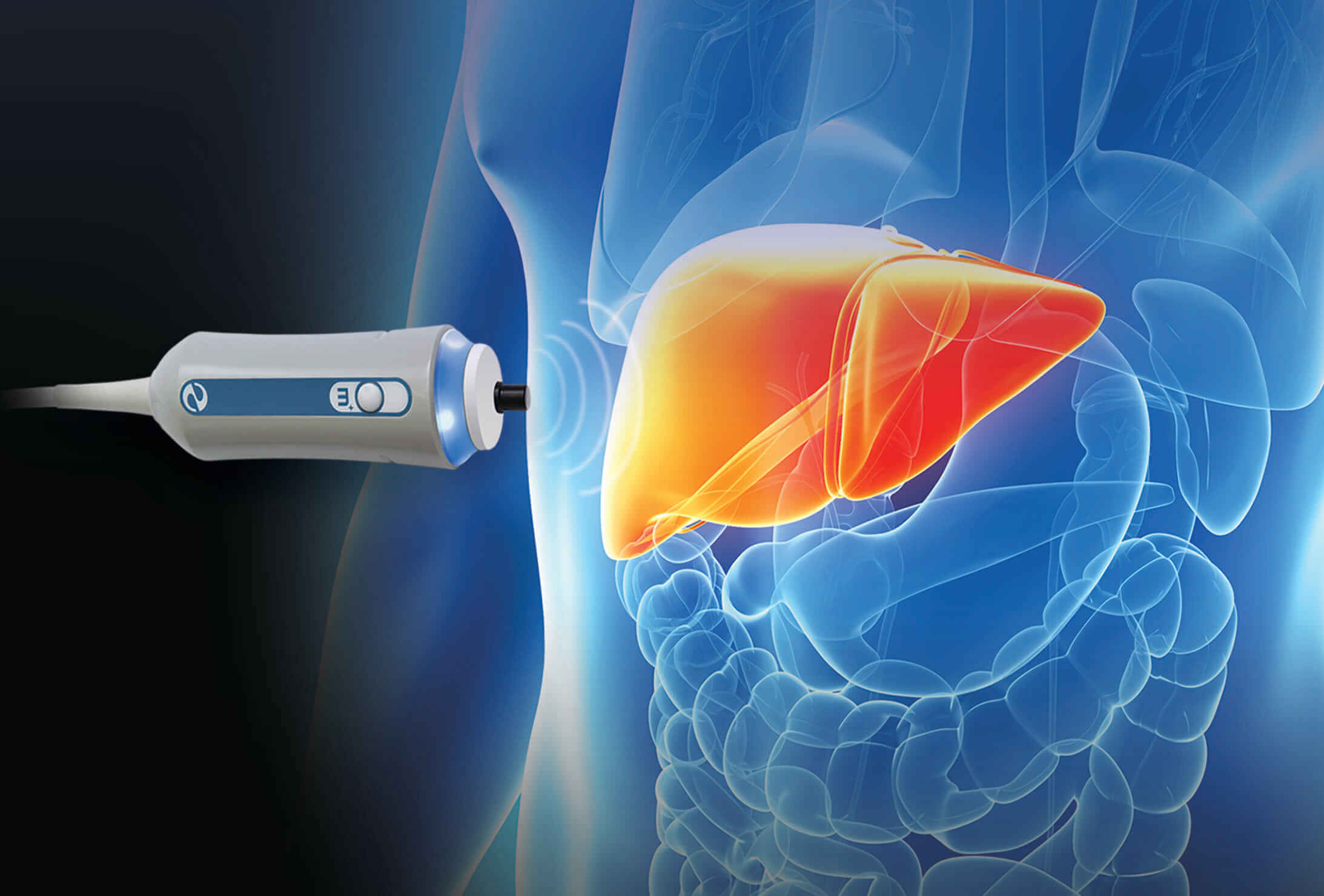
This is where FibroScan comes into play. It is a non‑invasive, accurate, and scientifically validated method that has changed physicians’ perspectives on diagnosing and staging fatty liver disease. Still, one fundamental question remains unanswered for many patients:
Is a FibroScan number just a number, or is it a serious warning about the future health of the liver?
This article is written precisely to clarify that uncertainty. In clear yet scientific language, we will explain what fatty liver disease actually is, how many stages it has, how FibroScan differs from ultrasound, and—most importantly—how to determine which stage you are currently in: a safe stage, or a point that requires serious action. If you have ever felt uncertain after receiving your ultrasound or FibroScan results, the rest of this article will help bring clarity.
What Exactly Is Fatty Liver Disease, and Why Is Staging It So Critical?
When we talk about fatty liver disease, we are referring to a broad disease spectrum, not a single, uniform condition. Non‑alcoholic fatty liver disease (NAFLD) refers to excessive fat accumulation within liver cells when alcohol consumption is not the primary cause. In many individuals, this condition can remain asymptomatic for years and may never progress to a dangerous stage.
But the story does not always end there. In a subset of patients, fatty liver disease enters a more active phase known as NASH (non‑alcoholic steatohepatitis)—a condition in which fat accumulation is accompanied by inflammation and liver cell injury. At this point, the disease moves beyond simple fat storage and enters the pathway of chronic liver damage. If this inflammation persists, the body attempts repair by forming fibrotic (abnormally stiff) tissue—a process known as liver fibrosis.
The critical point is this:
In fatty liver disease, the stage of the disease matters far more than the mere presence of fat. Two individuals may both have “fatty liver,” yet one may be in a completely reversible stage, while the other may be only one step away from cirrhosis. Without accurate staging, this crucial difference remains hidden.
The disease progression is usually slow and silent. Fibrosis develops gradually, without clear pain or obvious warning signs. That is why many patients only realize the seriousness of their condition when it has already reached advanced stages. Staging fatty liver disease is, in essence, a roadmap of your liver’s future—a map that shows where you stand and how much opportunity you still have to control the disease.
Why Ultrasound Alone Is Not Enough to Determine the Stage of Fatty Liver Disease
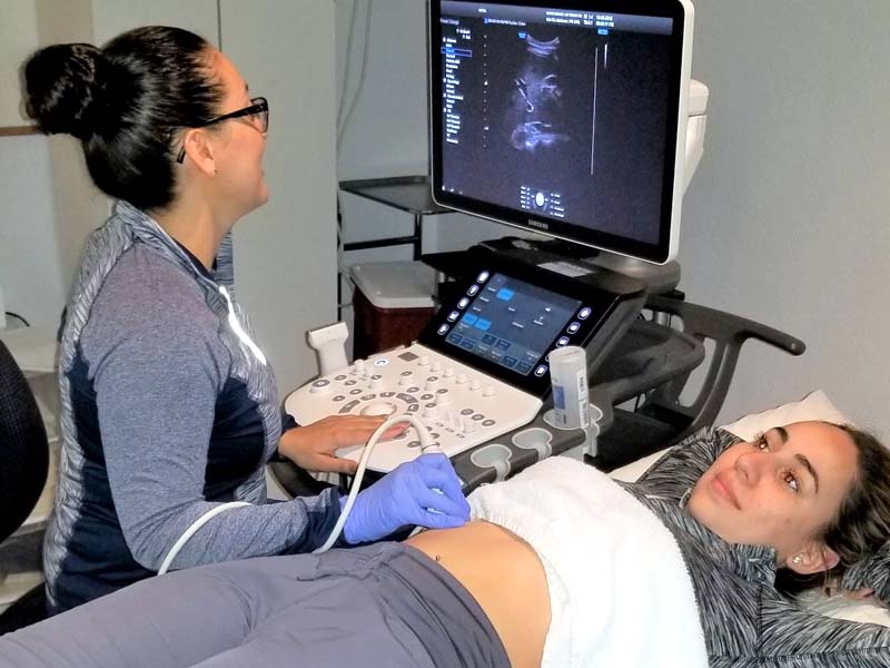
Ultrasound is the first and most accessible tool for diagnosing fatty liver disease—but its simplicity has led to a widespread misunderstanding. Many patients assume that ultrasound grading (grade 1, 2, or 3) directly reflects disease severity. In reality, ultrasound only shows the amount of fat accumulation, not the actual degree of liver tissue damage.
The main limitation is that ultrasound is not a reliable tool for detecting liver fibrosis. A person may have grade 1 fatty liver on ultrasound yet already have significant fibrosis; conversely, someone with important issue is what cannot be seen easily: the degree of liver stiffness and the possible onset of liver fibrosis.
This is where FibroScan comes into play. It is a non‑invasive, accurate, and scientifically validated method that has changed physicians’ perspectives on diagnosing and staging fatty liver disease. Still, one fundamental question remains unanswered for many patients:
Is a FibroScan number just a number, or is it a serious warning about the future health of the liver?
This article is written precisely to clarify that uncertainty. In clear yet scientific language, we will explain what fatty liver disease actually is, how many stages it has, how FibroScan differs from ultrasound, and—most importantly—how to determine which stage you are currently in: a safe stage, or a point that requires serious action. If you have ever felt uncertain after receiving your ultrasound or FibroScan results, the rest of this article will help bring clarity.
Complete Guide to Mammography: Why, How, and Key Tips Before Your Appointment
What Exactly Is Fatty Liver Disease, and Why Is Staging It So Critical?
When we talk about fatty liver disease, we are referring to a broad disease spectrum, not a single, uniform condition. Non‑alcoholic fatty liver disease (NAFLD) refers to excessive fat accumulation within liver cells when alcohol consumption is not the primary cause. In many individuals, this condition can remain asymptomatic for years and may never progress to a dangerous stage.
But the story does not always end there. In a subset of patients, fatty liver disease enters a more active phase known as NASH (non‑alcoholic steatohepatitis)—a condition in which fat accumulation is accompanied by inflammation and liver cell injury. At this point, the disease moves beyond simple fat storage and enters the pathway of chronic liver damage. If this inflammation persists, the body attempts repair by forming fibrotic (abnormally stiff) tissue—a process known as liver fibrosis.
The critical point is this:
In fatty liver disease, the stage of the disease matters far more than the mere presence of fat. Two individuals may both have “fatty liver,” yet one may be in a grade 2 or even grade 3 fatty liver may not have entered a serious structural damage phase. This discrepancy is a major source of error in treatment decisions.
In addition, the accuracy of ultrasound depends on many factors: the operator’s skill, the quality of the equipment, the patient’s body habitus, and even bowel gas. As a result, ultrasound grading is more of a descriptive imaging finding than a definitive measure of disease progression. This leads to an important question:
Does ultrasound truly reflect the severity of fatty liver disease?
The short answer is: not completely.
This is where the fundamental difference between ultrasound and FibroScan becomes clear. Instead of focusing solely on fat, FibroScan measures the actual stiffness of the liver—the factor that ultimately determines long‑term outcomes. For this reason, FibroScan has become the primary tool for scientific staging of fatty liver disease in recent years, with a role that extends far beyond simple ultrasound imaging.
In the next part of the article, we will examine in detail how FibroScan performs this staging and what each stage truly means for your liver health.
What Is FibroScan and How Does It Measure the Liver’s Actual Stiffness?
If we want to explain FibroScan in the simplest way, we should say that it is a tool that measures the true condition of the liver tissue instead of guessing. Unlike ultrasound, which mainly focuses on appearance and the amount of fat, FibroScan evaluates a much more important parameter: liver tissue stiffness. This stiffness is what determines whether your liver is on a healthy path of recovery or entering a phase of fibrosis and structural damage.
Scientifically speaking, FibroScan is a type of liver elastography. The device sends a gentle vibration wave into the liver tissue and measures the speed at which the wave returns. The stiffer the liver tissue is, the faster the wave comes back. The result is reported as a numerical value in kPa (kilopascals); a value that reflects the amount of tissue stiffness—not merely the presence of fat.
There is a common misconception here: many people think that a higher number automatically means “more severe fatty liver.” In reality, this number primarily reflects the degree of fibrotic change, not the amount of fat. Two people may have a similar amount of liver fat, yet their FibroScan numbers can be drastically different—and this difference completely changes their treatment path.
Compared to MRI, FibroScan is faster, more affordable, and far more practical for repeated follow‑up assessments. MRI provides excellent visual detail, but it is not always ideal for routine monitoring of disease progression. On the other hand, liver biopsy—although the diagnostic gold standard—is invasive, carries risks, and isn’t acceptable or necessary for all patients. FibroScan sits exactly in the middle: non‑invasive, accurate, and highly useful in clinical decision‑making.
For this reason, liver stiffness measurement has become one of the core foundations of diagnosing and staging fatty liver disease today.
Guide to Pregnancy Ultrasound Types and Their Clinical Applications
How Many Stages Does Fatty Liver Have on FibroScan? (Overview of Stages)
When you receive your FibroScan report, you often see terms like F0 to F4. These letters and numbers may appear confusing at first, but in reality they represent the roadmap of your liver’s disease progression. The important point is that FibroScan doesn’t just confirm fatty liver—it determines the stage.
Here’s what the staging means:
- F0: No fibrosis; the liver tissue still maintains its normal structure.
- F1: Very mild fibrosis has begun.
- F2: Moderate fibrosis—an important warning sign that treatment must be monitored.
- F3: Advanced fibrosis, considered close to cirrhosis.
- F4: Equivalent to liver cirrhosis; the liver’s natural architecture has undergone significant changes.
Here lies the key difference between simple fatty liver and liver fibrosis:
Fat accumulation can be temporary and reversible, but fibrosis is the result of ongoing inflammation and repeated improper tissue repair. This is why modern medicine focuses more on fibrosis stage than on fat content alone.
A crucial point is that the FibroScan number should not be memorized—it should be interpreted. Age, BMI, active inflammation, and even the time of day the test is performed can all influence the value. Therefore, FibroScan-based staging is not just a rigid classification; it is a tool for informed decision-making regarding treatment and preventing disease progression.
F0 and F1: Mild Fatty Liver | The Golden Window for Complete Recovery
When your FibroScan result falls within F0 or F1, there is genuinely good news hidden within it—provided that this opportunity is taken seriously. In these stages, the kPa value is usually low, indicating that the liver tissue has not yet developed significant stiffness. Put simply, the liver has not sustained meaningful structural damage, and full reversal is completely achievable.
In F0, there is essentially no fibrosis. The liver may be fatty, but its structure remains intact. In F1, extremely mild signs of fibrosis may be present—more of an early warning than permanent damage. This subtle difference underscores the importance of early detection.
One of the most frequently asked questions is:
Is F1 dangerous?
The short answer: No—not if it is not ignored.
F1 by itself is not dangerous, but if left unaddressed, it can progress. The difference between a controlled condition and a progressive disease is defined right at this stage.
In mild fatty liver with FibroScan results in this range, diet and lifestyle play a decisive role. Healthy weight loss, dietary modifications, increased physical activity, and proper management of blood sugar and lipids can not only stop progression but even return FibroScan values to a normal range. This stage is truly the golden window of treatment—a phase in which the disease can be fully reversed without strong medications or invasive procedures.
In the next sections, we will examine in detail what happens if the disease progresses beyond this stage—and why regular follow‑ups become critically important.
Stage F2: The Warning Point | The Beginning of True Liver Fibrosis
Stage F2 is where fatty liver disease is no longer just a simple metabolic disorder and begins to enter the phase of real structural liver damage. The key difference between F2 and F1 lies precisely here. In F1, fibrotic tissue is very mild and scattered, and the normal architecture of the liver is still preserved. In F2, however, fibrosis becomes more evident and more widespread, a stage physicians refer to as moderate liver fibrosis.
At this point, the liver’s repair processes no longer function in a fully healthy manner. Chronic inflammation causes liver cells, during regeneration, to produce stiff and abnormal tissue—what we refer to as fibrotic changes. These changes may not cause obvious daily symptoms, but they play a decisive role in determining the future course of the disease.
What is concerning is that many patients with F2 do not feel that they are at risk. They often have no significant pain and no clear warning signs that force them to seek follow‑up care. However, from a medical perspective, F2 is exactly the boundary between a disease that can be controlled primarily through lifestyle modification and one that requires serious medical monitoring. At this stage, general advice alone is no longer sufficient.
Regular follow‑up becomes critical. Periodic FibroScan assessments, liver function tests, and control of underlying factors such as diabetes, dyslipidemia, and excess weight can prevent progression to more dangerous stages of fatty liver disease. The good news is that F2 can still improve, but only if it is diagnosed in time and taken seriously.
Stages F3 and F4: When the Liver Enters a High‑Risk Phase
When FibroScan values reach the F3 or F4 range, the condition is no longer a silent or simple disease. These stages indicate that the liver has entered a high‑risk phase, and its natural structure has been significantly altered.
F3 is known as the pre‑cirrhotic stage. At this point, fibrosis has increased substantially, and stiff tissue involves a large portion of the liver. Normal blood flow through the liver becomes disrupted, and the liver’s functional reserve begins to decline. Nevertheless, some healthy tissue still remains. For this reason, disease control has not been completely lost—but the therapeutic window is far narrower.
In F4, however, the situation is fundamentally different. This stage is equivalent to liver cirrhosis, meaning that the normal liver architecture has been destroyed and replaced by fibrotic tissue. One of the most common questions at this stage is:
What does FibroScan F4 mean?
It means that the liver has largely lost its natural regenerative capacity, and the risk of complications such as liver failure, abdominal fluid accumulation (ascites), gastrointestinal bleeding, and even liver cancer increases significantly.
Is severe liver fibrosis treatable? The reality is that in F4, complete reversal is usually not possible. However, controlling disease progression, preventing complications, and preserving quality of life are entirely achievable. In F3, with targeted treatment and close follow‑up, a reduction in liver stiffness is sometimes even observed. The key difference lies in timing of diagnosis and the patient’s adherence to treatment.
Interpreting FibroScan Values (kPa): It’s Not Just a Number
One common mistake is judging FibroScan results solely based on the numerical value. In reality, a liver kPa value without proper clinical interpretation does not convey the full picture. While higher numbers generally indicate greater liver stiffness, this relationship is not always linear or straightforward.
Broadly speaking, lower values often correspond to F0–F1, intermediate values align with F2, and higher values trend toward F3 and F4. However, this is only a general framework—not an absolute rule. High BMI, active liver inflammation, congestion, and even eating before the test can falsely elevate FibroScan readings. This is why the same patient may show noticeably different results under different conditions.
This is where the physician’s role becomes essential. FibroScan results must be interpreted alongside blood tests, the patient’s medical history, and imaging findings. A kPa value may represent a serious warning for one patient, while for another it may simply indicate the need for routine follow‑up. Memorizing the number alone not only fails to help—it can create unnecessary anxiety.
In reality, FibroScan is a decision‑making tool, not a fear‑inducing one. When interpreted correctly, it helps determine the direction the liver is heading and identifies the right moment to intervene—before it becomes too late.
Can Fatty Liver Stages Be Controlled or Reversed?
This is the question nearly every patient asks:
Is fatty liver disease controllable, or does it inevitably get worse?
The honest answer is: yes, in many cases fatty liver disease can be controlled, and early stages can even be reversed—but not through exaggerated promises or commercialized “quick fixes.”
The first and most important pillar of treatment is genuine lifestyle modification. Not extreme short‑term diets, and not excessive exercise. Scientifically guided, gradual weight loss—typically 7–۱۰% of body weight—has been shown to directly reduce liver fat and improve liver fibrosis. Studies demonstrate that even this level of weight loss can lower FibroScan values.
Nutrition plays a central role. Reducing simple sugars, sweetened beverages, processed foods, and trans fats, while increasing fiber intake, healthy proteins, and beneficial fats, is one of the most effective ways to reverse fatty liver disease in F0 to F2 stages. These changes may seem modest, but their cumulative impact is substantial.
When it comes to medication, realism is essential. There is currently no drug that definitively cures liver fibrosis. However, in selected patients, medications aimed at controlling underlying conditions—such as diabetes, insulin resistance, or dyslipidemia—play an important supportive role. Medication is not a miracle cure, but when used appropriately and without exaggeration, it can help prevent disease progression.
Another critical factor is regular monitoring with FibroScan. Treating without monitoring is like driving without a speedometer. When FibroScan values begin to decline, it is a strong indicator that fatty liver disease is being managed correctly.
The Role of FibroScan in Treatment Monitoring
One of FibroScan’s greatest advantages is that it is not only a diagnostic tool—it is also a monitoring tool. It allows clinicians to assess whether treatment strategies are actually effective. Unlike blood tests, which can fluctuate, FibroScan provides a relative picture of structural changes within the liver.
A common patient question is:
How often should FibroScan be performed?
The answer depends on the disease stage. In mild stages (F0–F1), once a year or every 12–۱۸ months is usually sufficient. In F2 and above, follow‑up intervals are shorter—sometimes every 6–۹ months, depending on the patient’s condition and the severity of fibrotic changes.
The difference between pre‑ and post‑treatment values is highly meaningful. Even a reduction of 1–۲ kPa is considered a positive sign in many cases, indicating decreased inflammation and reduced liver stiffness. This helps physicians determine whether the current treatment approach is effective or needs adjustment.
Because of its ability to be repeated safely and frequently, FibroScan has, in many situations, largely replaced liver biopsy. Biopsy is invasive, risky, and unsuitable for repeated use. FibroScan, by contrast, is quick, painless, and repeatable—making it a cornerstone of long‑term fatty liver disease management.
Who Should Definitely Undergo a FibroScan?
Although FibroScan is not necessary for everyone, there are certain individuals who should not rely solely on ultrasound or blood tests. Being aware of this can prevent delayed diagnosis.
The first group includes people with diabetes, obesity, or high blood pressure. These three factors are among the strongest drivers of fatty liver disease progression and significantly increase the likelihood of moving from simple fat accumulation to fibrosis. In these individuals, even if the ultrasound findings are not very alarming, FibroScan is recommended.
The second group consists of individuals with a family history of liver disease or cirrhosis. Genetics plays a role in how quickly the disease progresses, and FibroScan helps clarify the liver’s true condition.
The third group includes those whose ALT or other liver enzymes remain persistently elevated, even when ultrasound shows only mild fatty liver. This mismatch between laboratory tests and imaging findings is an important warning sign.
Finally, addressing a very common question:
Who needs a FibroScan?
Any person with fatty liver disease who wants to know what stage their disease is in, whether it is reversible, or whether it requires serious medical follow‑up, is a reasonable candidate for undergoing FibroScan.
“FibroScan Is the Interpreter of Your Liver’s Future”
If we were to summarize the course of fatty liver disease in one sentence, it would be this:
The liver changes long before it starts to hurt.
From F0 and F1, where a golden opportunity for correction and complete reversal still exists, to F2, which is the warning point that sounds the alarm, and finally to F3 and F4, which indicate that the time for serious decisions has arrived.
In this journey, FibroScan plays the role of an “interpreter”—an interpreter that translates the silent language of the liver into numbers and understandable meaning. Not to scare, not to label, but to see the future path before it is too late. Knowing the stage of the disease means moving out of guesswork and into informed decision‑making.
The important point is that many patients, even with concerning numbers, still have the tools for control. Controlling fatty liver disease, reducing inflammation, and even improving liver fibrosis in early and intermediate stages is entirely possible—as long as the disease is recognized, not ignored. FibroScan makes this timely recognition possible.
This article is not meant to frighten you; it is meant to give you back the power of choice. The choice between ignorance and awareness, between waiting and follow‑up, between stress and control. Sometimes, a simple test can change the course of your liver’s health for years to come.
Knowing is the first step of treatment.
Frequently Asked Questions (FAQ)
Does a high FibroScan value always mean cirrhosis?
No. A high value does not necessarily mean cirrhosis. Factors such as inflammation, high BMI, liver congestion, or even test conditions can temporarily raise the number. Interpretation should always be done by a physician.
What is the difference between grade 2 fatty liver and F2?
The grade on ultrasound only reflects the amount of fat, while F2 on FibroScan indicates the degree of fibrosis (tissue stiffness). These are completely different concepts and cannot replace one another.
How often should FibroScan be repeated?
In mild stages, once a year is usually sufficient. In F2 and above, depending on the patient’s condition, it may be recommended every 6 to 9 months.
Is FibroScan painful?
No. FibroScan is completely painless, non‑invasive, and similar to a simple ultrasound. It takes only a few minutes.
How accurate is FibroScan?
It has high accuracy in detecting moderate to severe fibrosis, which is why it is used as a substitute for liver biopsy in many cases.
Does FibroScan value decrease with weight loss?
In many patients, yes. Scientific and gradual weight loss can reduce liver inflammation and stiffness, leading to an improvement in the kPa value.
Is FibroScan better than MRI?
Each has its own role. FibroScan is designed specifically for evaluating and monitoring fibrosis and is faster and more accessible, while MRI has broader diagnostic applications.
Is F3 reversible?
Complete reversal is not always possible, but in many cases control and even reduction of fibrosis can be achieved with proper treatment and follow‑up.
What is a normal FibroScan result (kPa)?
Generally, values below approximately 6–۷ kPa are considered normal or near‑normal, but precise interpretation depends on individual circumstances.
