Elastography
November 24, 2024
liver ultrasound
November 24, 2024Doctors use Doppler ultrasound to diagnose cardiovascular problems. This test shows the direction and speed of blood flow in the arteries and veins. Doppler ultrasound can identify blood clots, narrowed vessels, and other problems affecting the heart and blood vessels in the legs and arms, genital organs, chest, and thyroid. Doppler ultrasound is a type of ultrasound imaging test that uses sound waves to show the amount of blood flow in your blood vessels. It can be used to examine blood flow in many parts of the body, including various organs, the neck, arms, and legs.
Doppler ultrasound works by emitting sound waves to the red blood cells flowing in your veins. The ultrasound machine measures the echoes that return from the cells. Cells that move away from the sound waves produce different echoes compared to cells that move closer to the sound waves. This effect is called the Doppler effect. Doppler ultrasound is named in honor of Christian Doppler, a 19th-century physicist who discovered a method for measuring reflected sound waves from moving objects.

Types of Doppler ultrasound
Ultrasound is a non-invasive imaging test. A standard ultrasound typically produces images, but it does not show blood flow like a Doppler ultrasound.
Different types of Doppler ultrasound include:
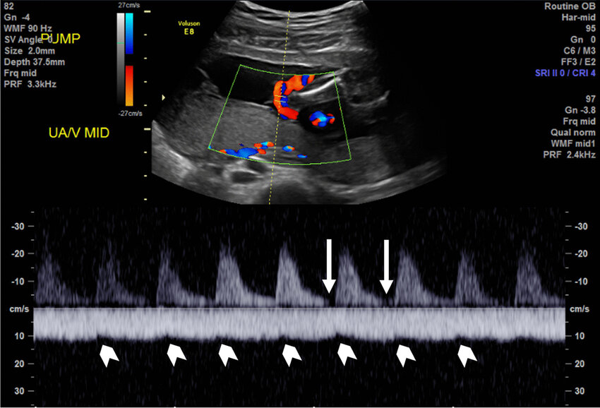
Color Doppler: A computer changes sound waves into different colors to show the direction of blood flow.
Spectral Doppler: A graphical display of blood flow over time.
Doppler ultrasound: Combines traditional ultrasound images with Doppler ultrasound. It can examine the width of blood vessels and help demonstrate vascular obstruction.
Power Doppler: This test is used to show the presence of blood flow and can be used to demonstrate very slow blood flow. It does not show the direction of blood flow. Providers may use power Doppler to study blood flow within the organs.
Transcranial Doppler ultrasound: Transcranial Doppler ultrasound examines blood flow in the brain to diagnose stroke or subarachnoid hemorrhage.
Some uses of Doppler ultrasound
Doctors use Doppler ultrasound for the following purposes:
Diagnose disorders affecting the blood vessels in the abdomen, legs, or arms.
After performing surgery or undergoing certain treatments and organ transplant surgery, they check the blood flow.
Evaluate the blood flow between the pregnant individual and the developing fetus during pregnancy.
Doctors use Doppler ultrasound to diagnose the following:
Narrowing of arteries or veins.
Blood clots, including deep vein thrombosis (DVT).
Damage to blood vessels.
Chronic venous insufficiency (CVI).
Assessment of blood supply to the transplanted organ (such as the kidney, liver, or pancreas).
Causes of renal vascular hypertension.
Tumor in the blood vessels.

How should I prepare for a Doppler ultrasound?
Depending on the type of ultrasound and the reason for the test, you may need to:
Fast for a few hours before the test (do not eat or drink).
Quit smoking and do not use nicotine products at least two hours before the test. Nicotine constricts blood vessels, which may affect the test results.
How a Doppler ultrasound is performed
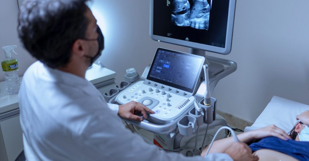
Doppler ultrasound is often performed by a sonography specialist who has received special training to conduct ultrasound examinations. Depending on the part of the body being examined, the test may be performed in various ways. Most Doppler ultrasound examinations include these general steps:
You remove your clothing from the area being examined and lie down on the table.
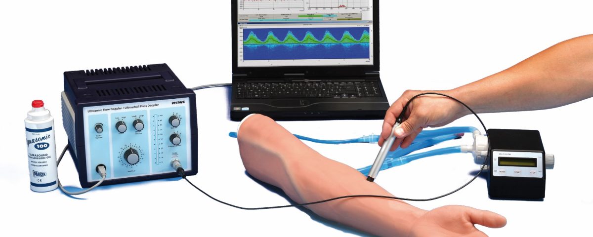
The specialist applies a special gel to the area of your skin that is being examined.
The specialist moves a wand-like device called a transducer over your skin. This device sends sound waves into your body, but you cannot feel them.
Sound waves are reflected off the red blood cells in your bloodstream.
When the ultrasound machine captures the echoes and converts them into an image or graph on the computer screen, you may hear sounds.
After the examination is over, the technician will wipe the gel off your body.
For some ultrasound examinations, the ultrasound device is placed inside the body cavity. Depending on the organs being examined, the device may be placed in the following cases:
Vagina: This is called a transvaginal ultrasound, which helps in observing the uterus and ovaries.
Rectum: This is called a transrectal ultrasound, which is usually performed to observe the prostate gland.
Esophagus (the organ that connects your mouth and stomach): This type of echocardiogram is called transesophageal echocardiogram (TEE). This is done to obtain clear images of the heart.

What are the risks of Doppler ultrasound?
Doppler ultrasound is a non-invasive and low-risk test. There is no need for injected contrast dyes like in angiography or the use of radiation like X-rays or CT scans. Ultrasound is not harmful or painful. It is safe enough for providers to use them on a pregnant individual.
Interpretation of Doppler ultrasound results
If the Doppler ultrasound results are normal, it means that the blood vessels that were examined appear to be healthy. They are not narrowed or have blood clots, and the blood flow is normal. If your results are not normal, the meaning depends on the area of the body and the type of Doppler ultrasound you have undergone. Talk to your doctor about what your results mean for your health and the need for treatment.


