
breast biopsy
May 18, 2025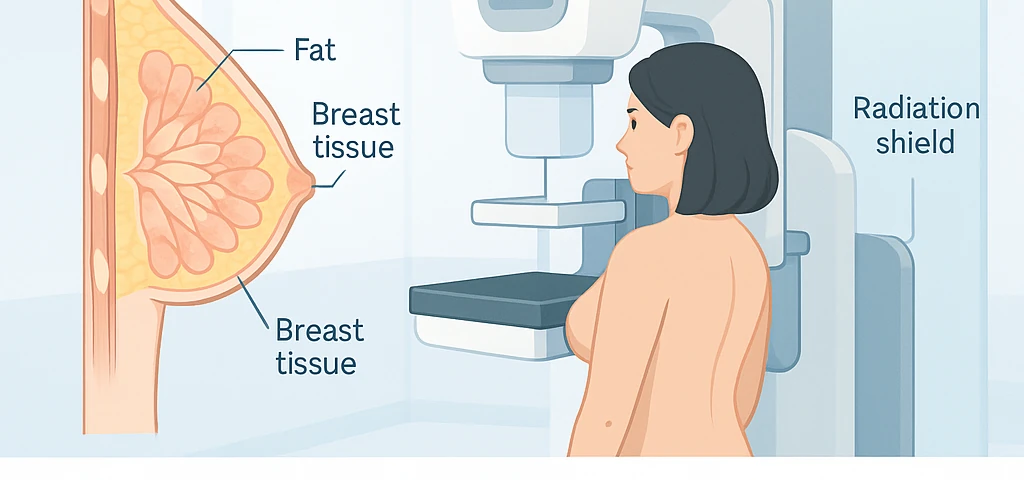
The Truth About Mammography
Mammography is one of the most vital tools for the early detection of breast cancer—a condition that is highly treatable in its initial stages. However, when diagnosis is delayed, the risk of metastasis and treatment complications increases.
Over the past decade, statistical analyses by the Ministry of Health have shown that early detection through mammography can increase the likelihood of successful treatment by up to 90%.
In this path, the existence of specialized imaging centers equipped with modern technology—such as the Golestan Radiology and Sonography Center—has become increasingly important. Using full‑field digital systems, this center enables physicians to observe the finest tissue details, preventing delays or errors in diagnosis and helping patients gain peace of mind.
Golestan Radiology and Sonography Center
The Golestan Radiology and Sonography Center has been operating since 2004, under the management of Dr. Marjan Zare (Radiologist) and Dr. Abdolreza Sajadian (Interventional Radiology Subspecialist). Relying on extensive expertise and long‑standing experience in breast imaging, the center is recognized as one of the most trusted institutions for patients in Shiraz and southern Iran.
Advanced Technologies and Equipment
All imaging equipment at Golestan Center meets international quality standards, including:
Full‑Field Digital Mammography (FFDM): The most advanced breast imaging technology, providing ultra‑high resolution with minimal radiation dose.
Shear Wave Elastography: An innovative technique for evaluating breast tissue stiffness and differentiating benign from malignant lesions.
Digital Radiography (DDR): A rapid digital system for general and specialized imaging with a significant reduction in radiation exposure.
Commitment to Women’s Health
Golestan Center follows all international women’s health protocols. Its calm and safe environment, combined with the supervision of an experienced team, creates a comfortable and reassuring experience for every patient.
Online Access and Consultation
On the official website of the center, separate sections are dedicated to Golestan Mammography Services, Breast Ultrasound Report Interpretation, and Breast Elastography. Patients can access specialized information and schedule their appointments online with ease.
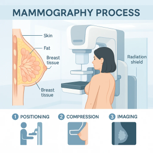
What Is Mammography and How Is It Performed?
Mammography is one of the most important medical imaging techniques that uses low-dose X-rays to display the internal structure of breast tissue. Its main purpose is the early detection of abnormalities before the appearance of visible symptoms such as lumps or skin changes.
At the Golestan Radiology and Sonography Center, full‑field digital mammography systems are used to capture high‑resolution images of both breasts from multiple angles. Compared with older models, this technology offers greater accuracy, faster performance, and lower radiation exposure.
The mammography procedure is remarkably simple: the patient stands in front of the device, the breast is gently placed between two transparent plates, and a detailed image is captured within seconds. This brief compression ensures clearer imaging and causes no harm to the tissue.
For women with dense breast tissue or younger patients, breast ultrasound is performed as a complementary method to improve diagnostic accuracy. Combining these techniques at Golestan Radiology Center enables specialists to detect even small or hidden lesions with precision.
Mammography at this center is accurate, fast, safe, and completely standardized, providing a comfortable and pain‑free experience that reassures patients about their health and well‑being.

Types of Mammography and Their Differences
Mammography methods are generally divided into three main categories based on their purpose and the type of technology used. The physician selects the most appropriate type according to the patient’s condition.
۱٫ Screening Mammography:
This type is recommended for women over 40 years of age or those with a family history of breast cancer. Its main objective is to identify very small changes in breast tissue before a palpable lump or visible symptom appears.
۲٫ Diagnostic Mammography:
When a patient presents with symptoms such as a palpable lump, pain, or abnormal nipple discharge, the physician performs this type to closely examine the suspicious areas. It is often done after screening or as a second‑stage evaluation.
۳٫ Digital 3D Mammography (Tomosynthesis):
In this advanced technique, the device takes multiple images of the breast from different angles and reconstructs them into thin layers, providing a three‑dimensional view. This technology significantly increases the chance of detecting small and early‑stage tumors, especially in women with dense breast tissue.
At the Golestan Radiology and Sonography Center, the latest Full‑Field Digital Mammography (FFDM) systems are used. These systems automatically compare previous and current images, allowing tissue changes as small as a few millimeters to be clearly identified.
How Mammography Images Are Interpreted
After imaging, the results are reviewed by a breast radiology specialist and reported based on the BI‑RADS system (Breast Imaging Reporting and Data System). The goal of this system is to standardize the description of findings and determine the likelihood of malignancy.
The radiologist carefully examines image details, evaluating tissue density, presence of masses, microcalcifications (tiny calcium deposits), and any suspicious skin or nipple changes. The final result is then categorized according to BI‑RADS, which serves as a universal language among doctors and imaging centers.
What Do BI‑RADS Categories 0–۶ Mean?
BI‑RADS 0: Incomplete – additional imaging (such as ultrasound) is required.
BI‑RADS 1 & 2: Negative or benign findings.
BI‑RADS 3: Probably benign; follow‑up in 6 months is recommended.
BI‑RADS 4: Suspicious abnormality; biopsy suggested.
BI‑RADS 5: Highly suggestive of malignancy (greater than 95% likelihood).
BI‑RADS 6: Proven malignancy confirmed through biopsy.
At the Golestan Radiology and Sonography Center, patients can receive clear, patient‑friendly explanations of their mammography reports. If necessary, they may also request specialized interpretation services from the Golestan Mammography Interpretation Department for more precise analysis of results.
Warning Signs of Breast Changes You Should Never Ignore
Every woman should be familiar with warning symptoms that may indicate breast cancer or other abnormal breast changes. The most important signs include:
- Presence or detection of a lump in the breast or underarm
- Noticeable change in size or shape of one breast compared to the other
- Skin dimpling, puckering, or changes in the nipple’s shape or texture
- Unusual nipple discharge (especially bloody or watery)
- Persistent localized pain, redness, or swelling of the skin
If you experience any of these symptoms, you should visit a specialized imaging center such as Golestan Radiology and Sonography Center as soon as possible. Through mammography and complementary ultrasound, the cause can be accurately identified and, if necessary, early treatment initiated.
Important Tips Before Mammography
Proper preparation plays a crucial role in obtaining clear and accurate images. The following recommendations are essential:
- The best time for mammography is one week after menstruation, when the breast tissue is softer and less tender.
- Avoid using deodorants, powders, perfumes, or lotions on your chest or underarms on the day of your exam, as these products may appear as calcium deposits on images.
- If you are pregnant, breastfeeding, or have undergone previous breast surgery, inform your physician or radiology technician.
- Bring your previous mammography or ultrasound images so your doctor can compare them and evaluate any subtle changes.
At Golestan Center, all procedures are carried out with high precision, full privacy assurance, and supervised by specialized physicians to ensure patient comfort and trust.
Is Mammography Painful or Dangerous?
During mammography, the breast is gently compressed between two plates for a few seconds to obtain a clear image. This brief pressure may cause mild discomfort, but it does not harm breast tissue.
The advanced digital systems at Golestan Radiology and Sonography Center allow precise adjustment of compression and radiation levels, ensuring maximum image clarity with minimal discomfort.
The radiation dose used is significantly lower than international safety limits, with no long‑term risks to health.
Therefore, mammography performed at Golestan Center is a safe, quick, and accurate method that provides women with reassurance and confidence in their breast health.
The Importance of Combining Mammography, Ultrasound, and Elastography
In younger women and particularly in those with dense breast tissue, mammography alone may not always detect all abnormalities. For this reason, physicians recommend performing breast ultrasound as a complementary method to provide a more accurate evaluation of dense areas.
Alongside these two imaging techniques, the advanced Shear Wave Elastography technology serves as a third diagnostic step. This method measures the stiffness of breast tissue, helping physicians distinguish between benign (softer) and malignant (firmer) lesions with high precision.
At the Golestan Radiology and Sonography Center, all three diagnostic technologies—digital mammography, breast ultrasound, and shear wave elastography—are available in one integrated facility.
Patients can complete all necessary evaluations in a single short session, which not only saves time but also enhances diagnostic accuracy. Each imaging record is digitally stored, allowing for easy comparison in future follow‑up visits, ensuring consistency and accuracy in patient monitoring.
Limitations and Possible Errors in Mammography
Although mammography is one of the most effective screening tools for early breast cancer detection, it still has certain limitations. In women with dense breast tissue, X‑rays may not fully penetrate the tissue, which can lead to hidden or unrecognized lesions. The presence of breast implants may also reduce image clarity and diagnostic accuracy.
Other factors such as patient movement during imaging, improper positioning of the breast, or low cooperation levels may cause image blurring and potential misinterpretation.
In such cases, the radiologist may recommend re‑imaging or additional methods such as breast MRI to obtain a more comprehensive assessment.
At the Golestan Radiology and Sonography Center, all imaging procedures are performed under the direct supervision of specialists, minimizing both technical errors and the need for repeat imaging.
Post‑Mammography Care and Follow‑Up Recommendations
If the mammography report reveals an abnormality or suspicious area, your physician may recommend one or more of the following steps:
- Targeted breast ultrasound or fine‑needle biopsy for tissue examination
- Short‑term follow‑up imaging (every 3 to 6 months) for monitoring subtle changes
- Referral to a breast surgeon for final diagnostic evaluation, if a lesion is confirmed
For women aged 40 and older, annual mammography remains the most effective method to prevent delayed diagnosis of breast cancer.
The Golestan Center also offers a comprehensive breast‑health monitoring program, allowing patients to undergo regular evaluations and maintain an electronic record of previous images. This enables physicians to track progression and compare results at each visit.
According to Dr. Marjan Zare, radiologist, and Dr. Abdolreza Sajadian, interventional radiology subspecialist,
“Prevention is always easier and more effective than treatment.”
Timely and precise mammography not only saves lives but also simplifies the treatment process and reduces overall costs.
Through the integration of digital mammography and shear‑wave elastography, Golestan Center has introduced a new standard in breast cancer screening, combining accuracy, comfort, and efficiency for every patient.
Frequently Asked Questions (FAQ)
۱٫ How often should mammography be performed?
Women aged 40 and above are advised to undergo mammography once a year.
In those with a family history of breast cancer, shorter intervals—every 6 to 12 months—may be recommended.
۲٫ Is mammography possible during breastfeeding?
During lactation, the breast tissue is filled with milk glands, which can reduce image clarity.
However, if a lump or clinical symptom is present, low‑dose mammography can still be safely performed under medical supervision.
۳٫ What is the difference between mammography and breast ultrasound?
Mammography uses low‑dose X‑rays to identify changes in fat and tissue density, while ultrasound uses sound waves and is better suited for examining dense tissue or cystic lesions.
۴٫ Is mammography necessary for younger women?
Women under 40 who have a family history of breast cancer or suspicious symptoms should consult their physician regarding the need for mammography or ultrasound.
۵٫ What do BI‑RADS results mean?
The BI‑RADS system is an international reporting standard that classifies findings from 0 to 6, ranging from normal to confirmed malignancy.
۶٫ How much radiation does mammography involve?
The radiation dose in modern mammography is significantly below international safety limits and poses no harm to the patient’s health.
۷٫ Can mammography and elastography be performed together at Golestan Center?
Yes. At the Golestan Radiology and Sonography Center, mammography, breast ultrasound, and shear‑wave elastography can all be performed in a single coordinated session, ensuring that the final diagnostic report is delivered to the patient as quickly and accurately as possible.
