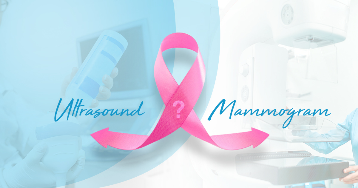
The difference between mammography and ultrasound
October 8, 2024
Complete Guide to Mammography: Why, How, and Key Tips Before Your Appointment
November 4, 2025
A breast biopsy is the process of removing a sample of breast tissue for evaluation. The sample of breast tissue will be sent to a laboratory. Your doctor may recommend a breast biopsy if you have some suspicious area in your breast (such as a breast lump or other signs and symptoms suggesting breast cancer). The results of a breast biopsy can tell whether the suspicious area is breast cancer. Each pathology report from a breast biopsy can aid in determining whether you are a candidate for additional surgery or another treatment option.
What is a breast biopsy?
A breast biopsy is when a doctor removes a sample of your breast tissue to look at under a microscope. A breast biopsy is (to date) the only way a doctor can absolutely rule out breast cancer. You may be feeling nervous or scared about having a breast biopsy. Though it is completely understandable, you should always remember that having a breast biopsy does not mean you have breast cancer. In fact, 80 percent of women who have a breast biopsy do not have breast cancer.
Reasons for having a breast biopsy
There is a lump or thickening in the breast you or your doctor can feel, and your doctor believes you may have breast cancer.
There is suspicious area in the breast noted in a mammogram.
There is abnormal finding on an ultrasound or magnetic resonance imaging (MRI) of the breast.
There are abnormal changes to the nipple or areola (the dark colored skin surrounding the nipple). These changes include flaking of the skin, indentation of the skin, or other abnormal discharge.
Types of breast biopsy
There are several types of breast biopsy and there are different approaches for the different types. The following are the types:
Fine Needle Aspiration Procedure (FNA): This is a procedure that is performed by a radiologist using a syringe with a thin needle to acquire fluid and/or tissue from the breast.
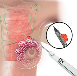
Core needle breast biopsy: This type of biopsy is performed by radiologists. A core needle breast biopsy is performed using a larger needle than that used in an FNA; since the needle is larger, the radiologist is able to obtain more breast tissue than that acquired by FNA.
A breast excisional biopsy (lumpectomy): This procedure is done to remove all the potentially cancerous tissue in the area identified by tests to have abnormalities. The surgeon may remove some surrounding normal tissue to also confirm that all cancerous cells have been removed.
An incisional breast biopsy: The surgeon only removes a portion of the abnormal tissue in the area of interest.
The risks and complications of breast biopsy
The risks associated breast biopsy include:
Bruising of the breast, with swelling of the breast.
Infection or bleeding at the biopsy site.
Change in the appearance of the breast, depending on the size of the tissue that was removed and healing of the breast.
Need to have further surgery or other treatments, as determined by the biopsies results.
If you develop a fever, if the biopsy site becomes red or warm, or you have any unusual discharge from the biopsy site, please call your healthcare team. This may be a sign of infection that needs to be treated as soon as possible.
How to prepare for your breast biopsy
First, if you have any of the following experiences, please notify your doctor:
You’re pregnant or possibly pregnant.
You are taking blood thinners (anti-coagulants).
You’re taking aspirin or herbal products.
Having any allergies, especially to anesthesia
Having any illness or had surgery in past year.
If you cannot lie on your stomach for an extended period of time.
If you have a pacemaker or any other electronic device implanted in your body, let your doctor know.
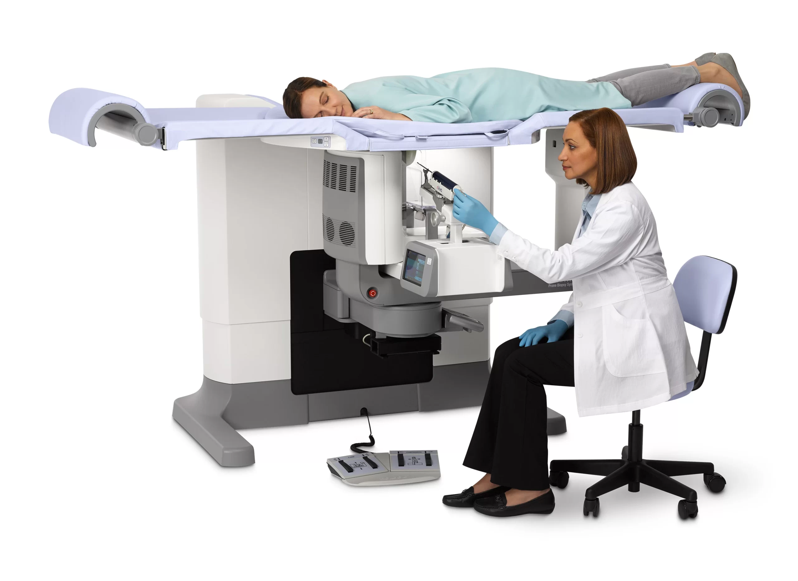
What is to be expected during a breast biopsy?
Your experience will vary depending on the breast biopsy that is performed by your physician:
If you have a fine needle aspiration biopsy the doctor will perform a local anesthetic. A needle is inserted into your skin to retrieve fluid and tissue.
If you have a central needle breast biopsy, your radiologist will give you a local anesthetic. Depending on what the radiologist will do, they may make a small incision in your breast in order to insert the needle which will be used to take the tissue sample, and they will place biopsy markers in the biopsy area. The markers are used in case tissue specimens are required to examine the biopsy area again.
If you are having surgery (such as a lumpectomy or excisional biopsy), your anesthesiologist will first put you to sleep. The surgeon will make an incision(s) in your breast to remove tissue samples. The incisions will be closed with sutures and covered with a sterile bandage.
How long does a breast biopsy take?
How long a procedure takes depends on what kind of biopsy it is.
A fine needle aspiration may take 10 to 15 minutes.
A core needle breast biopsy takes approximately 20 to 60 minutes, depending on the imaging process your radiologist takes to guide them.
A breast biopsy (lumpectomy) generally takes up to 60 minutes.
A breast excisional biopsy generally takes approximately 60 minutes.
After a breast biopsy
All breast biopsy types except surgical biopsy allow you to go home with only a bandage and an ice pack applied to the biopsy site. Most women want to rest for the rest of the day after a breast biopsy but can return to normal activities in approximately a day.
Bruising is common after a core needle biopsy. You may take a non-aspirin pain medication (containing acetaminophen – Tylenol, Excedrin) to decrease pain and discomfort after a breast biopsy and apply cold pack on swelling, if needed.
If you have had a surgical biopsy, you probably have some stitches. You will likely go home the same day of your surgery, and usually can resume your normal activities the next day. The health care team will let you know how to care for your stitches.
Breast biopsy results
It may take a few days before the breast biopsy results are ready. After the biopsy has been done, the breast tissue is sent to the lab. The lab has a specialist in blood and tissue examination (a pathologist) check the specimen under a microscope and by using other methods.
The pathologist produces a pathology report that goes to your doctor. Your doctor will review the report with you. The pathology report describes the size and characteristics of the tissue specimens and the biopsy area. It describes if there were cancer, benign changes, or precancerous cells noted.
If the pathology report states only healthy tissue (or benign changes in the breast) have been diagnosed, your doctor should check to see if the radiologist and pathologists concur. The conclusions by these two experts occasionally may vary. For example, the radiologist may see that your mammogram had a more ominous-looking lesion, something like breast cancer or precancerous lesion but your pathology report reflects only normal breast tissue. In this case, you may require further surgery to provide additional tissue for examination of the area.
If the pathology report states that there is breast cancer present, it will describe the cancer, that is the type of breast cancer you have, and provide further details, such as whether the hormone receptors are positive or negative. At that point, you and your doctor can make a treatment plan that works best for you and fits your needs.