
What is an anomaly ultrasound?
February 1, 2025
nuchal translucency (NT) ultrasonography
August 23, 2025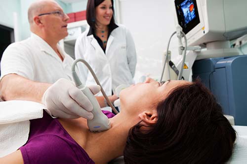
When evaluating a mass or nodule located in the neck, one of the primary things that doctors want to rule out is the presence of cancer, but they may also wish to know about the function of your thyroid gland. Ultrasound is an imaging technique that uses high-frequency sound waves to create images of the thyroid gland. Ultrasound provides the best detail about the shape and structure of nodules as well as the ability to determine whether a nodule is solid or cystic or if there are multiple nodules. Additionally, doctors sometimes use ultrasound to guide a fine needle aspiration biopsy.
As we mentioned, ultrasound uses sound waves to produce images of the thyroid gland in the neck. There is little forethought, medical preparation, or preparation required. Jewelry should not be brought along, and you should wear loose, comfortable clothing for the appointment. You may be instructed to wear a gown for the ultrasound.
What exactly is a thyroid ultrasound?
Ultrasound imaging is a kind of non-invasive medical test frequently used to help doctors identify and treat medical conditions. The method is safe and painless, and uses sound waves to produce images from inside the body. To use ultrasound, a small probe against the skin (called a transducer) and some gel are used. The probe is placed directly against the skin, and the gel helps the sound waves travel through the skin. The transducer transmits and receives the high frequency sound waves through the gel and into your body. The transducer picks up the reflected sound waves and a computer uses the reflected sound waves to build a picture. X-rays are not used for ultrasound exams. Because ultrasound captures moving images, it depicts the structure and movement of your body’s internal organs. The pictures obtained from ultrasound imaging can also be used to determine blood flow in blood vessels.
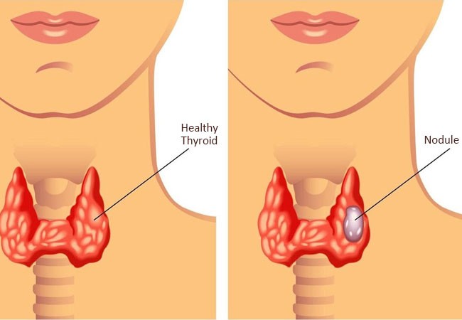
A thyroid ultrasound is a special ultrasound that makes images of the thyroid gland and surrounding tissues in the neck. The thyroid is located in the front of the neck directly under the Adam’s apple (or laryngeal prominence) and is shaped like a butterfly. The thyroid is also one of nine endocrine glands located throughout the body that produce hormones that are sent directly into the bloodstream.
The thyroid synthesizes thyroid hormone, which aids in the regulation of several functions in the body, including heart rate. The development of thyroid nodules or spots, sometimes palpable within the skin, is exceedingly common. Approximately 5 to 10 percent of adults have identifiable nodules in their thyroid that a doctor can feel during a physical examination. Ultrasound is very sensitive and detects many more nodules that can only be viewed and not palpated. For instance, in some age categories, nodules are observed on ultrasound in 70 percent of adults. The vast majority of these areas are benign thyroid tissue, they are not health threats. In a few instances, they are thyroid tumors that may need more diagnosis and treatment.

Some typical purposes of thyroid ultrasound are:
- Determine if a mass in the neck is from the thyroid, or from structures close to the thyroid.
- Examine the characteristics of thyroid nodules to determine if they are benign and common nodules, or if the nodule has characteristics that require a biopsy. In the event a biopsy is required, fine needle aspiration guided by ultrasound may provide better accuracy for the biopsy.
- Evaluate for additional nodules in patients with one or more palpable nodules by physical examination.
To check how thyroid nodules have changed, for example how much growth there has been over time.
Because ultrasound is live imaging, a doctor may use it to help guide procedures like fine needle biopsies. In biopsies, a needle will take a tissue sample to the lab. They also utilize ultrasound to help guide placement of drainage, like catheters.
For a thyroid ultrasound how should you prepare?
Dress comfortably in loose-fitting clothing. You will be asked to remove any jewelry in the area that will be imaged. You may be asked to wear a gown.
There are no other preparations necessary.
It has to be emphasized that ultrasound examinations are very sensitive to motion, therefore an active or crying child will increase the length of the examination. If your child is calm, it will go much more smoothly. To keep children calm, some parents bring a book, a small toy, music or some games so that the child can focus on those things to help the specialist do their job effectively.
The equipment used for a thyroid ultrasound and how the procedure is performed
Ultrasound machines consist of a computer console, video monitor, and a transducer attached by an electric cord. The transducer is a small, handheld device, which looks somewhat like a microphone. The transducer emits high-frequency sound waves that are inaudible to humans into the body. The transducer listens for the echoes returning to the transducer.
The physician applies a small amount of gel to the area under examination and places the transducer on the gel. The gel provides an interface for the sound waves to travel back and forth from the transducer to the area under examination. The ultrasound image is provided in real-time, on the video monitor. The computer creates the ultrasound image based on the amplitude (range), pitch (frequency) and time it takes for the ultrasound signal to return to the transducer. The computer also considers the type of body structure or tissue the sound is traveling through.
Usually, there is no discomfort or pressure, however, if the area of interest is sensitive you may feel slight pressure or pain caused by the transducer. Once imaging is complete, the technician asks the Patient does the technician will wipe off the clear ultrasound gel from your skin. Any remaining ultrasound gel will dry very quickly. Ultrasound gel typically does not stain or discolor clothing.
What will I experience during and after a thyroid ultrasound?
Most ultrasound examinations are painless, quick, and easily tolerable. A thyroid ultrasound usually takes about 30 minutes.
If you are suffering from neck pain, inform the technician so they can help you get into a suitable position for the examination.
Benefits versus risks of thyroid ultrasound
Advantages:
Most ultrasound scans are non-invasive (without needles or injections).
Sometimes, an ultrasound examination may be temporarily uncomfortable, but it shouldn’t be painful.
Ultrasound is widely available, easy to use, and cheaper than many other imaging methods.
Ultrasound imaging is very safe and does not use radiation.
An ultrasound scan provides a clear image of soft tissues that are not well shown in X-ray images.
Ultrasound provides real-time and live imaging. This makes it a good tool for guiding minimally invasive procedures such as needle biopsy and fluid aspiration.
Risks:
Standard diagnostic ultrasound has no known harmful effects on humans.
After the ultrasound examination, you can immediately resume your normal activities.
The function of the thyroid cannot be assessed with an ultrasound which means whether the gland is underactive, overactive, or normal. The physician may order a blood test, or radioactive iodine uptake test, for this diagnosis. The physician may order a blood test, or radioactive iodine uptake test, for this diagnosis.